The cell cycle is a fundamental biological process that governs cellular replication, growth, and genomic integrity. It is essential for normal development and physiology, but its disruption is closely linked to disease, especially cancer. In oncology, analyzing the cell cycle is crucial for understanding and predicting tumor behavior. Cancer often involves defects in cell cycle regulation. Dysregulation of cell cycle checkpoints and signaling pathways is widely regarded as an initiating event in carcinogenesis, contributing to uncontrolled proliferation and genomic instability. Tracking changes in cell cycle progression also helps researchers assess how drugs work, measure their effectiveness, and uncover potential resistance mechanisms.
In this article, we highlight recent methods of assessing the cell cycle in mammalian cells, with a focus on commonly used molecular cell cycle markers. The cell cycle can be briefly summarized as consisting of four key phases: G1 (gap 1), S (DNA synthesis), G2 (gap 2), and M (mitosis). Cells that exit the cell cycle, such as various differentiated cells, may enter a quiescent state known as G0. These phases are tightly controlled by the expression of key proteins, including cyclin-dependent kinases (CDKs), cyclins, CDK inhibitors, tumor suppressors, and transcription factors. Key markers associated with each cell cycle phase are described below.
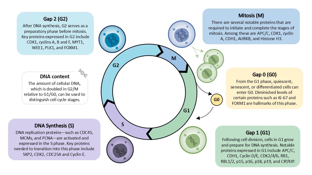
This diagram highlights the phases of the cell cycle and commonly associated markers.
G1 phase markers
Cells complete mitosis and enter the G1 phase with the help of the anaphase-promoting complex (APC/C), activated by CDH1. This activation drives the continued degradation of mitotic and S-phase cyclins, preventing premature entry into S phase. During G1, cells grow in size until they receive mitogenic signals that trigger pathways for S-phase entry. Among these signals, D-type cyclins (D1, D2, D3) are upregulated and bind to CDK4 or CDK6. The resulting active complexes phosphorylate RB1, RBL1, and RBL2, leading to the release and activation of E2F transcription factors. For most of G1, RB1 remains mono-phosphorylated, becoming fully phosphorylated by cyclin E–CDK2 complexes at the G1/S transition. Cyclin E–CDK2 also inactivates the APC/C–CDH1 complex, facilitating progression to promote the transition into the S phase.
On the other hand, the INK4 family of CDK inhibitors (p15, p16, p18, p19) suppresses CDK4/6 activity, maintaining the G1 state. The CIP/KIP family of inhibitors further restrains CDK2, preventing premature entry into the S-phase. In the event of DNA damage, CHK2 and p53 will become active, leading to CDK downregulation and arrest at the G1/S transition.
S phase markers
Cellular DNA is replicated in the S phase, a tightly regulated and irreversible process. CDK inhibitors are degraded by regulatory proteins such as SKP2 to promote the G1/S phase transition. To further complete the transition, CDK2 is activated by phosphatases (such as CDC25A) and binds with cyclin E. Upon entering the S phase, cyclin A2 accumulates and complexes with CDK2 to phosphorylate components of the DNA replication machinery. Key proteins required to initiate DNA replication include CDC45, the MCM helicase complex, and the PCNA sliding clamp. The amount of cellular DNA is doubled upon the completion of the S phase.
G2 phase markers
The G2 phase is a preparatory stage before mitosis, in which the cell grows and prepares for division. During this phase, cyclin F levels peak, playing several regulatory roles: it suppresses E2F activity to halt further DNA replication, inhibits the synthesis of replicative histones, regulates centrosome duplication, and targets CDH1 for degradation. Cyclin A remains active and initiates the activation of the cyclin B-CDK1 complex, a key driver of mitotic entry. However, CDK1 activation is tightly regulated. Phosphorylation by the CAK complex and dephosphorylation by CDC25B and CDC25C are required for full activation, while inhibitory kinases MYT1 and WEE1 counteract this process to prevent premature mitosis.
Cyclin B (B1 and B2), initially confined to the cytoplasm during G2, must be phosphorylated by PLK1 to translocate into the nucleus, where it can activate CDK1 and initiate chromosomal segregation. Aurora A kinase, which accumulates at centrosomes from S phase onward, is crucial for centrosome maturation and spindle formation. It also phosphorylates PLK1, further promoting CDK1 activation, spindle formation, and progression into mitosis. The transcription factor FOXM1 binds to promoters containing CHR elements, activating the transcription of genes essential for mitotic entry and progression.
M phase markers
When the cell meets the requirements of the G2 checkpoint, it enters the M phase of the cell cycle. At the onset of mitosis, cyclin B accumulates in the nucleus and forms a complex with CDK1, which phosphorylates various target proteins to drive mitotic events, including spindle formation, chromosome condensation, and nuclear envelope breakdown. APC/C is a key regulator here, facilitating the metaphase-to-anaphase transition by targeting specific proteins for degradation. Cyclin A is among its first substrates degraded after nuclear envelope breakdown and before metaphase. Later, APC/C mediates the degradation of securin and cyclin B, which triggers sister chromatid separation and the onset of anaphase.
Antibodies Search Tool Search Now Find and compare marker-specific antibodies across leading reagent suppliers.
As mitosis progresses, CDK1 activity declines, enabling chromosome segregation and the completion of mitosis and cytokinesis. During anaphase, APC/C in association with CDH1 ensures the degradation of remaining mitotic regulators, including Aurora A and PLK1, to allow proper exit from mitosis. The aurora B kinase, a key component of the chromosomal passenger complex, localizes to chromosomes and the mitotic spindle, playing essential roles in chromosome condensation, alignment, and cytokinesis. Notably, Aurora B phosphorylates histone H3 on serine 10, a modification that serves as a hallmark of mitotic chromatin and a widely used marker for identifying mitotic cells.
G0 phase markers
For a variety of reasons, such as limited nutrients or a fully differentiated state, cells may exit the cell cycle and enter the non-replicative G0 state. Cells in the G0 phase can be further classified as quiescent, senescent, or differentiated. In contrast to the other cell cycle phases, markers of the G0 phase often involve assessing for a deficiency of certain proteins or genetic material. The well-known proliferation marker Ki-67 is present in all stages of the cell cycle and faces degradation when cells complete division in G1. In non-dividing G0 cells, Ki-67 is absent. Another example is the downregulation of the transcription factor FOXM1, which marks quiescent and non-proliferative cells. RNA content, believed to be lower in content in a resting or quiescent state compared to the G1 phase, can be assessed by pyronin Y staining.
DNA as a cell cycle marker
Measuring cellular DNA content has been a widely used method to distinguish different cell cycle phases. Cells in the G1 and G0 phases contain a diploid amount of DNA. This is half the DNA content found in cells in the G2 and M phases, in which DNA replication has been completed. This difference in DNA content can be detected through the use of DNA-binding fluorescent dyes such as propidium iodide, DAPI, or Hoechst, to distinguish the distinct cell cycle phases of cell populations via flow cytometry or fluorescence microscopy. Many commercial cell cycle assay kits also utilize this mechanism as a rapid method of cell cycle determination.
While this method effectively identifies G1/G0 and G2/M cells, it cannot distinguish the intermediate levels of actively replicating DNA in the S phase. For S phase detection, nucleotide analogs such as 5-bromo-2′-deoxyuridine (BrdU) and 5-ethynyl-2′-deoxyuridine (EdU) can be incorporated into newly synthesized DNA during replication and later detected with antibodies or chemical reactions.
DNA-based assessment of the cell cycle, while ideal for fast and routine applications, can face some limitations. They provide only static and relative data about the length of distinct phases. DNA labeling allows for kinetic measurements, but also introduces some toxicity that should be taken into account for longer-term studies.
Table of cell cycle markers
The table below lists markers of cell cycle phases as mentioned by recent literature. Marker information is accompanied by links to relevant antibody and ELISA kit products, as these are commonly used in marker immunodetection. The associated products are offered by a variety of manufacturers and, when used in combination with other markers, can serve as a useful reference for characterizing cells in the various stages of the cell cycle.
| Marker | Gene | Marker Type | Molecule Type | Description | Reference | Antibodies | ELISA Kits |
|---|
| APC/C |
- |
M, G1 |
Protein Complex |
Controls mitotic exit by degrading Cyclin B and Securin |
1,3,4 |
|
|
| Aurora A |
AURKA |
G2/M, Cancer |
Kinase |
Activates CDC25B and spindle formation |
2,4,5 |
AURKA antibodies |
AURKA ELISA |
| Aurora B |
AURKB |
M, Cancer |
Kinase |
Controls chromosome segregation and cytokinesis |
2,7 |
Aurora B Kinase antibodies |
Aurora B Kinase ELISA |
| BrdU |
- |
S |
DNA Label |
DNA synthesis markers used to detect cells in S phase |
6,7 |
BrdU antibodies |
BrdU ELISA |
| CAK |
CDK7, CCNH, MNAT1 |
Other |
Protein Complex |
Activates CDKs via phosphorylation |
2,4 |
CAK antibodies |
CAK ELISA |
| CDC6 |
CDC6 |
G1 |
DNA-Binding Protein |
Primes origins for DNA replication |
4 |
Cdc6 antibodies |
Cdc6 ELISA |
| CDC45 |
CDC45 |
G1/S |
DNA-Binding Protein |
Initiates DNA replication, loading onto chromatin after CDK activation |
4 |
Cdc45 antibodies |
Cdc45 ELISA |
| CDC25A |
CDC25A |
G1/S, Cancer |
Phosphatase |
Activates CDK2 for S phase entry |
2,4,5 |
CDC25A antibodies |
CDC25A ELISA |
| CDC25B |
CDC25B |
G2/M, Cancer |
Phosphatase |
Activates CDK1 for mitotic entry |
5 |
CDC25B antibodies |
CDC25B ELISA |
| CDC25C |
CDC25C |
G2/M, Cancer |
Phosphatase |
Activates CDK1 for mitotic entry |
2,5 |
CDC25C antibodies |
CDC25C ELISA |
| CDH1 |
FZR1 |
G1 |
Ubiquitin Ligase |
Activates APC/C to prevent premature entry into S phase |
3,4 |
CDH1 antibodies |
CDH1 ELISA |
| CDK1 |
CDK1 |
G2/M, Cancer |
Kinase |
Essential for mitotic entry |
1-6 |
CDK1 antibodies |
CDK1 ELISA |
| CDK2 |
CDK2 |
G1/S, S, Cancer |
Kinase |
Promotes DNA replication and S phase |
1-6 |
CDK2 antibodies |
CDK2 ELISA |
| CDK3 |
CDK3 |
G0/G1, G1/S |
Kinase |
Promotes G0/G1 and G1/S transition |
1,3 |
|
|
| CDK4 |
CDK4 |
G1, Cancer |
Kinase |
Phosphorylates Rb to promote G1/S transition |
1-7 |
CDK4 antibodies |
CDK4 ELISA |
| CDK5 |
CDK5 |
Cancer |
Kinase |
Plays non-canonical roles in neuronal cells |
1,2 |
CDK5 antibodies |
CDK5 ELISA |
| CDK6 |
CDK6 |
G1, Cancer |
Kinase |
Phosphorylates Rb to promote G1/S transition |
1-6 |
CDK6 antibodies |
CDK6 ELISA |
| CDK7 |
CDK7 |
Other, Cancer |
Kinase |
Activates CDKs |
1,2,4 |
|
|
| CDK9 |
CDK9 |
Other, Cancer |
Kinase |
Regulates transcription |
2,4 |
CDK9 antibodies |
CDK9 ELISA |
| CDT1 |
CDT1 |
G1 |
DNA-Binding Protein |
Primes origins for DNA replication; FUCCI component |
4,7 |
CDT1 antibodies |
CDT1 ELISA |
| CHK1 |
CHEK1 |
S, G2/M, Cancer |
Kinase |
Initiates DNA damage checkpoints |
2,5 |
Chk1 antibodies |
Chk1 ELISA |
| CHK2 |
CHEK2 |
G1 |
Kinase |
Initiates DNA damage checkpoints |
1,2 |
|
|
| c-Myc |
MYC |
G1 |
Transcription Factor |
Drives cell cycle progression via cyclin/CDK expression |
2,3,4 |
c-Myc antibodies |
c-Myc ELISA |
| Cyclin A2 |
CCNA2 |
S, G2, Cancer |
Binding Protein |
Controls S phase and entry into mitosis |
1,2,3,5 |
Cyclin A2 antibodies |
Cyclin A2 ELISA |
| Cyclin B1 |
CCNB1 |
G2/M, Cancer |
Binding Protein |
Partners with CDK1 for mitosis |
2,7 |
Cyclin B1 antibodies |
Cyclin B1 ELISA |
| Cyclin B2 |
CCNB2 |
G2/M, Cancer |
Binding Protein |
Partners with CDK1 for mitosis |
2,5 |
Cyclin B2 antibodies |
Cyclin B2 ELISA |
| Cyclin D1 |
CCND1 |
G1, Cancer |
Binding Protein |
Activates CDK4/6 to promote G1/S transition |
1,2,3,4 |
Cyclin D1 antibodies |
Cyclin D1 ELISA |
| Cyclin D2 |
CCND2 |
G1, Cancer |
Binding Protein |
Activates CDK4/6 to promote G1/S transition |
2,3,4 |
Cyclin D2 antibodies |
Cyclin D2 ELISA |
| Cyclin D3 |
CCND3 |
G1, Cancer |
Binding Protein |
Activates CDK4/6 to promote G1/S transition |
1,2,3,4 |
CCND3 antibodies |
CCND3 ELISA |
| Cyclin E1 |
CCNE1 |
G1/S, Cancer |
Binding Protein |
Partners with CDK2 for S phase entry |
1-5 |
Cyclin E antibodies |
Cyclin E ELISA |
| Cyclin E2 |
CCNE2 |
G1/S, Cancer |
Binding Protein |
Partners with CDK2 for S phase entry |
2,3,4,5 |
CCNE2 antibodies |
CCNE2 ELISA |
| Cyclin F |
CCNF |
G2 |
Binding Protein |
Regulates degradation of proteins involved in DNA repair |
1,4 |
CCNF antibodies |
CCNF ELISA |
| Cyclin H |
CCNH |
Other, Cancer |
Binding Protein |
Component of CAK complex |
2,4 |
CCNH antibodies |
CCNH ELISA |
| DAPI |
- |
G1, S, G2, M |
DNA Stain |
Fluorescent DNA stain |
6 |
|
|
| E2F Family Proteins |
E2F1–8 |
G1/S |
Protein Family |
Transcription factor driving expression of S-phase genes |
2,3,4 |
E2F antibodies |
E2F ELISA |
| E2F1 |
E2F1 |
G1/S |
Transcription Factor |
Transcription factor driving expression of S-phase genes |
1,5 |
E2F1 antibodies |
E2F1 ELISA |
| EdU |
- |
S |
DNA Label |
DNA synthesis markers used to detect cells in S phase |
6,7 |
|
|
| FOXM1 |
FOXM1 |
G2/M, G0 |
Transcription Factor |
Regulates expression of mitotic genes |
2,5 |
FOXM1 antibodies |
FOXM1 ELISA |
| Geminin |
GMNN |
S, G2, M |
Binding Protein |
Inhibits DNA re-replication by suppressing Cdt1; FUCCI component |
6,7 |
Geminin antibodies |
Geminin ELISA |
| Ki-67 |
MKI67 |
G1, S, G2, M, G0 |
DNA-Binding Protein |
Marker of proliferation, absent in G0 |
1,6 |
Ki-67 antibodies |
Ki-67 ELISA |
| MCM |
MCM2–7 |
G1/S |
Protein Complex |
Licensing and initiation of DNA replication |
2,4,5 |
|
|
| MYT1 |
PKMYT1 |
G2 |
Kinase |
Inhibits CDK1 to delay mitosis |
2,4 |
Myt1 antibodies |
Myt1 ELISA |
| ORC |
ORC1–ORC6 |
G1 |
Protein Complex |
Binds origins of replication for licensing |
4 |
ORC antibodies |
ORC ELISA |
| p15 |
CDKN2B |
G1 |
Binding Protein |
CDK inhibitors, especially targeting CDK4/6 |
1,2,3,4 |
CDKN2B antibodies |
CDKN2B ELISA |
| p16 |
CDKN2A |
G1 |
Binding Protein |
CDK inhibitors, especially targeting CDK4/6 |
1,2,3,4 |
CDKN2A antibodies |
CDKN2A ELISA |
| p18 |
CDKN2C |
G1 |
Binding Protein |
CDK inhibitors, especially targeting CDK4/6 |
1,2,3,4 |
p18 antibodies |
p18 ELISA |
| p19 |
CDKN2D |
G1 |
Binding Protein |
CDK inhibitors, especially targeting CDK4/6 |
1,2,3,4 |
CDKN2D antibodies |
CDKN2D ELISA |
| p21 (CIP1) |
CDKN1A |
G1/S |
Binding Protein |
CDK inhibitor, regulates cell cycle arrest |
1,2,3,4 |
CDKN1A antibodies |
CDKN1A ELISA |
| p27 (KIP1) |
CDKN1B |
G1/S |
Binding Protein |
Inhibit CDK activity to control S phase entry |
1,2,3,4 |
CDKN1B antibodies |
CDKN1B ELISA |
| p53 |
TP53 |
G1/S, G2/M |
Transcription Factor |
Responds to DNA damage by activating p21 |
1,2,7 |
p53 antibodies |
p53 ELISA |
| p57 (KIP2) |
CDKN1C |
G1/S |
Binding Protein |
Inhibits CDK activity to control S phase entry |
2,3,4 |
CDKN1C antibodies |
CDKN1C ELISA |
| PCNA |
PCNA |
S |
DNA-Binding Protein |
Sliding clamp for DNA polymerase; replication marker |
1,6,7 |
PCNA antibodies |
PCNA ELISA |
| p-Histone H3 S10 |
- |
M |
Phosphorylated Protein |
Marker of mitosis and chromosome condensation |
6,7 |
|
|
| p-RB |
RB1 |
G1/S |
Phosphorylated Protein |
Inactivated Rb promotes E2F release |
1,7 |
|
|
| PLK1 |
PLK1 |
G2/M, Cancer |
Kinase |
Promotes mitotic entry and spindle function |
2,4 |
PLK1 antibodies |
PLK1 ELISA |
| Propidium iodide |
- |
G1, S, G2, M |
DNA Stain |
Fluorescent DNA stain |
6 |
|
|
| Pyronin Y |
- |
G0 |
RNA Stain |
Fluorescent RNA stain |
6 |
|
|
| RB1 |
RB1 |
G1 |
Binding Protein |
Suppresses E2F to block S-phase gene expression |
2,3,4 |
RB1 antibodies |
RB1 ELISA |
| RBL1 |
RBL1 |
G1 |
Binding Protein |
Suppresses E2F to block S-phase gene expression |
3,4 |
RBL1 antibodies |
RBL1 ELISA |
| RBL2 |
RBL2 |
G1 |
Binding Protein |
Suppresses E2F to block S-phase gene expression |
3,4 |
RBL2 antibodies |
RBL2 ELISA |
| Skp2 |
SKP2 |
G1/S |
Ubiquitin Ligase |
Degrades p27 to promote S phase entry |
3,4 |
SKP2 antibodies |
SKP2 ELISA |
| WEE1 |
WEE1 |
G2, Cancer |
Kinase |
Inhibits CDK1 to prevent premature mitosis |
2,4,5 |
WEE1 antibodies |
WEE1 ELISA |
References
Sharma R, Kumar D, Jha NK, Jha SK, Ambasta RK, Kumar P. Re-expression of cell cycle markers in aged neurons and muscles: Whether cells should divide or die? Biochim Biophys Acta Mol Basis Dis. 2017 Jan;1863(1):324-336. doi: 10.1016/j.bbadis.2016.09.010. Epub 2016 Sep 14. PMID: 27639832.
Otto T, Sicinski P. Cell cycle proteins as promising targets in cancer therapy. Nat Rev Cancer. 2017;17(2):93-115. doi:10.1038/nrc.2016.138
Liu L, Michowski W, Kolodziejczyk A, Sicinski P. The cell cycle in stem cell proliferation, pluripotency and differentiation. Nat Cell Biol. 2019;21(9):1060-1067. doi:10.1038/s41556-019-0384-4
Suski JM, Braun M, Strmiska V, Sicinski P. Targeting cell-cycle machinery in cancer. Cancer Cell. 2021;39(6):759-778. doi:10.1016/j.ccell.2021.03.010
Riba, A., Oravecz, A., Durik, M. et al. Cell cycle gene regulation dynamics revealed by RNA velocity and deep-learning. Nat Commun 13, 2865 (2022). https://doi.org/10.1038/s41467-022-30545-8
Ligasová A, Frydrych I, Koberna K. Basic Methods of Cell Cycle Analysis. Int J Mol Sci. 2023;24(4):3674. Published 2023 Feb 12. doi:10.3390/ijms24043674
Chen YL, Chen YC, Suzuki A. ImmunoCellCycle-ID: A high-precision immunofluorescence-based method for cell cycle identification. Preprint. bioRxiv. 2024;2024.08.14.607961. Published 2024 Aug 15. doi:10.1101/2024.08.14.607961