Autophagy is an intracellular pathway for delivering everything from proteins, to organelles, to pathogens, to the lysosome for degradation and recycling. It is thought of as the cell’s waste disposal system and problems in its functioning have downstream ramifications. “Autophagy has critical roles in cell signalling, development, and immunity, which is exemplified by mouse knock-out models that show loss of autophagy leads to various pathologies, including tumorigenesis and neurodegeneration,” says Ian Ganley, Ph.D., Programme Leader, School of Life Sciences, University of Dundee. Several studies have indeed confirmed the role of autophagy in the onset and development of cancer, neurodegeneration, autoimmunity, infection, and inflammation.
Complex nature of autophagy
As autophagy connects many cellular events, modulating it could potentially eliminate the need to separately target multiple pathways. However, targeting autophagy is not that simple because there are different types of autophagy and each involves organelles and cellular pathways that are not well understood. Autophagy is also a dynamic process, and the steady state levels of autophagosomes or autophagic proteins do not necessarily indicate what’s happening in the pathway as a whole, in terms of enhancement or inhibition. “It’s all about the flux,” says Ganley. “More autophagosomes does not necessarily mean more autophagy, as a similar situation could arise if lysosomal trafficking/degradation is impaired—and hence autophagy is blocked.”
The autophagic response is also strictly context dependent. For instance, in neurodegenerative aggreopathies, such as Huntington’s and Alzheimer’s disease, autophagy activation has been shown to aid in clearance of aggregated protein. Whereas in cancer, autophagy inhibition is sometimes thought to be beneficial as autophagy increases cell survival. “The impairment of the autophagic flux can promote or counteract tumorigenesis, depending on the cancer type and stage,” explains Rosaria Esposito, Ph.D., Application Scientist at Enzo Life Sciences. “We, therefore, need integrative approaches to profile autophagy and identify novel markers and regulatory factors to develop precision therapeutics.”
Jean Mulcahy-Levy, M.D., Associate Professor and Investigator in the Morgan Adam’s Foundation Pediatric Brain Tumor Research Program at the Children's Hospital at the University of Colorado in Denver, agrees that targeting autophagy is all about balance. “In cancer we have found a window of availability where we can block autophagy and not cause any of the side effects that you might see. Sometimes, it is good to activate autophagy to get rid of any abnormal cells to prevent cancer, and if you get the cancer you switch to blocking autophagy to kill the tumor cells. You have to take each situation in context and then choose the right path for intervention.” Hence, both activators and inhibitors of autophagy are being developed for therapeutic use, depending on the biological need.
Figuring out the when and where
Autophagy is a multi-step process and some of the new drugs that are in preclinical development target ULK1 or Vps34 proteins that are at the beginning of the autophagy pathway. There are other drugs in development that build on the effectiveness of existing autophagy inhibitors, like chloroquine and hydroxychloroquine, to make them work better. “We often wonder whether it’s better to target the start or the end of the autophagy process, or somewhere in between,” says Mulcahy-Levy.
Although hydroxychloroquine does inhibit autophagy and mTORC1 inhibitors activate autophagy, they both do so indirectly and have many autophagy-independent effects. “It would be helpful to have direct, specific inhibitors and activators of the autophagy pathway for use in vivo and eventually in clinical trials,” says Malia Potts, Ph.D., Senior Scientist, Oncology Research at Amgen. Since autophagy encompasses many different pathways with conflicting biological outcomes, there is a need to find precise tools and robust biomarkers to monitor and characterize the distinct pathways. “While the core autophagy machinery is well-understood, at present we do not have the necessary pharmacological tools to manipulate it effectively in patients. Add to this the complexities of regulatory mechanisms controlling each step and the existence of multiple forms of selective autophagy,” says Potts.
A promising new approach to exploit autophagy for therapeutic purposes involves chemically targeting the autophagy machinery to induce degradation of a target protein. “With autophagy targeting chimeras (AUTACs), the idea is that it may be possible to degrade previously “undruggable” targets by pharmacologically linking them to the autophagy-lysosome pathway,” says Potts. Proteolysis targeting chimeras (PROTACs) have already been shown to successfully hijack the ubiquitin-proteasome system, for targeted protein degradation and are now in clinical development.
Subham Basu, Ph.D., Director of Strategy for Immuno-oncology at Abcam, says that having methodologies to select the right targets and design the PROTAC for specific tissues and diseases, and having tools to monitor the degradation in vivo will be critical for this work. “Regardless of which aspect of autophagy one is working on—upstream regulation, mechanisms of the process itself, downstream effects—or whether it’s basic, translational, or clinical research, having the right model system and tools is vital. This includes antibodies that work for the relevant applications, including in vivo, as well as the right cell lines and biochemical reagents.”
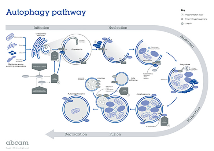
Image: Overview of the autophagy process. An expanding membrane structure (phagophore) enwraps portions of the cytoplasm, followed by the formation of a double-membrane sequestering vesicle (autophagosome). The autophagosome fuses with the lysosome and releases its inner compartment into the lysosomal lumen. The inner membrane part of the autophagosome is degraded together with the enclosed cargo. The resulting macromolecules are released into the cytosol for recycling through lysosomal membrane permeases. Image courtesy of Abcam.
Overcoming the current limitations
One of the limitations in the study of autophagy has been trying to capture the dynamic process with static measurements and trying to draw biological inferences from them. “For example, the expression levels of typical markers used to monitor the autophagy process, such as LC3 or p62, can change independent of the progression of the autophagic flux itself,” says Esposito. “Therefore, the availability of a unique marker or a standard methodology, allowing the unambiguous interpretation of the results is needed.”
Another concern in autophagy has been the assigning of cellular function based on the presence or absence of markers, in a given physiological setting. “In any given cell type, depending on the stimulus or target, it is possible that the rate of autophagosome formation may be slower than their rate of degradation, hence there may be an accumulation of autophagosomes or vice versa,” says Ganley. “It is therefore very important to determine the amount of autophagic protein that is being lysosomally degraded, i.e. the flux.”
Finally, although the overall autophagy processes are well mapped out, there is still a lot of work to be done to figure out how the different components of the pathway interact with each other, especially when designing drugs that specifically target a certain protein or gene. A key aspect to understanding how and when to therapeutically target autophagy will involve determining not only when and where it becomes activated in vivo, but also what is specifically being degraded. “The advent of fluorescent autophagy reporters to monitor specific autophagy pathways such as mitophagy, pexophagy, etc. and their applications in creating transgenic mouse models will be critical in this endeavor,” says Ganley. There are several commercially available dyes, antibodies, recombinant proteins, knock-out cell lines, and assays for detecting various aspects and components of autophagy, either individually or as a panel of markers.
Taking into account the complexity of the autophagic process, and the inherent limitations in the methodologies for capturing this dynamic process, the use of multiple approaches and markers, and the inclusion of proper controls in the experiment is highly recommended for reducing the risk of data misinterpretations, says Esposito. “When using drugs to induce or inhibit autophagy, one should use the minimal required concentration and use short incubation times in order to avoid off-target effects. A cell viability test can also be included as a control during the experiment, as these compounds may likely be toxic and trigger apoptosis or other signaling pathways.”
Mulcahy-Levy whose work focuses on studying autophagy in pediatric brain tumors uses tools such as Western blotting, flow cytometry, and electron microscopy to measure levels of autophagy. She uses different disease models, such as cells, tumor-bearing mice, and other model organisms, to study autophagy-induced cellular changes. “We collaborate with our clinical trial specialists to get samples from the patient if they are being treated with autophagy-altering drugs. This gives us an opportunity to specifically study how these drugs are changing the genetic profile, metabolism, or cell survival in these patients.”
Hero image from Abcam. Fluorescent microscopy showing nucleus (blue, DAPI) and autophagic vesicles (green, FITC) in HepG2 cells treated with 0.1 mM chloroquine for 24 hours. In earlier stages of autophagy, chloroquine induces autophagosome formation.