Western blot gel electrophoresis
In the Western blot workflow, protein gel electrophoresis follows sample preparation. The procedure is necessary for transforming the complex protein mixture in the sample into a structured, spatially resolved array that enables the distinction of individual proteins. The separation is most commonly carried out using polyacrylamide gel electrophoresis under denaturing conditions, which relies on the sodium dodecyl sulfate detergent (SDS-PAGE). SDS binds proteins along their backbone, denaturing and linearizing them while imparting a uniform negative charge proportional to their mass. Under an applied electric field, proteins migrate through the polyacrylamide matrix at rates inversely related to their size, with smaller proteins traveling more quickly through the pores. The result is a separation determined by molecular mass rather than native charge or conformation. Proteins can also be resolved under native conditions (native PAGE), which preserves protein structure and interactions, though this approach is far less common in Western blotting workflows.
Electrophoresis also serves as an important quality control checkpoint for the experiment. Running a prestained molecular weight marker alongside experimental samples provides a size reference, allowing users to confirm that detected bands correspond to the expected molecular weight of the target protein. The banding patterns visible in the gel after the run can also be used to assess whether the protein was loaded uniformly across lanes. A well-executed electrophoretic separation is therefore critical for generating reproducible, interpretable Western blot data. As SDS-PAGE is by far the most widely used separation method, its general procedure is discussed in detail below.
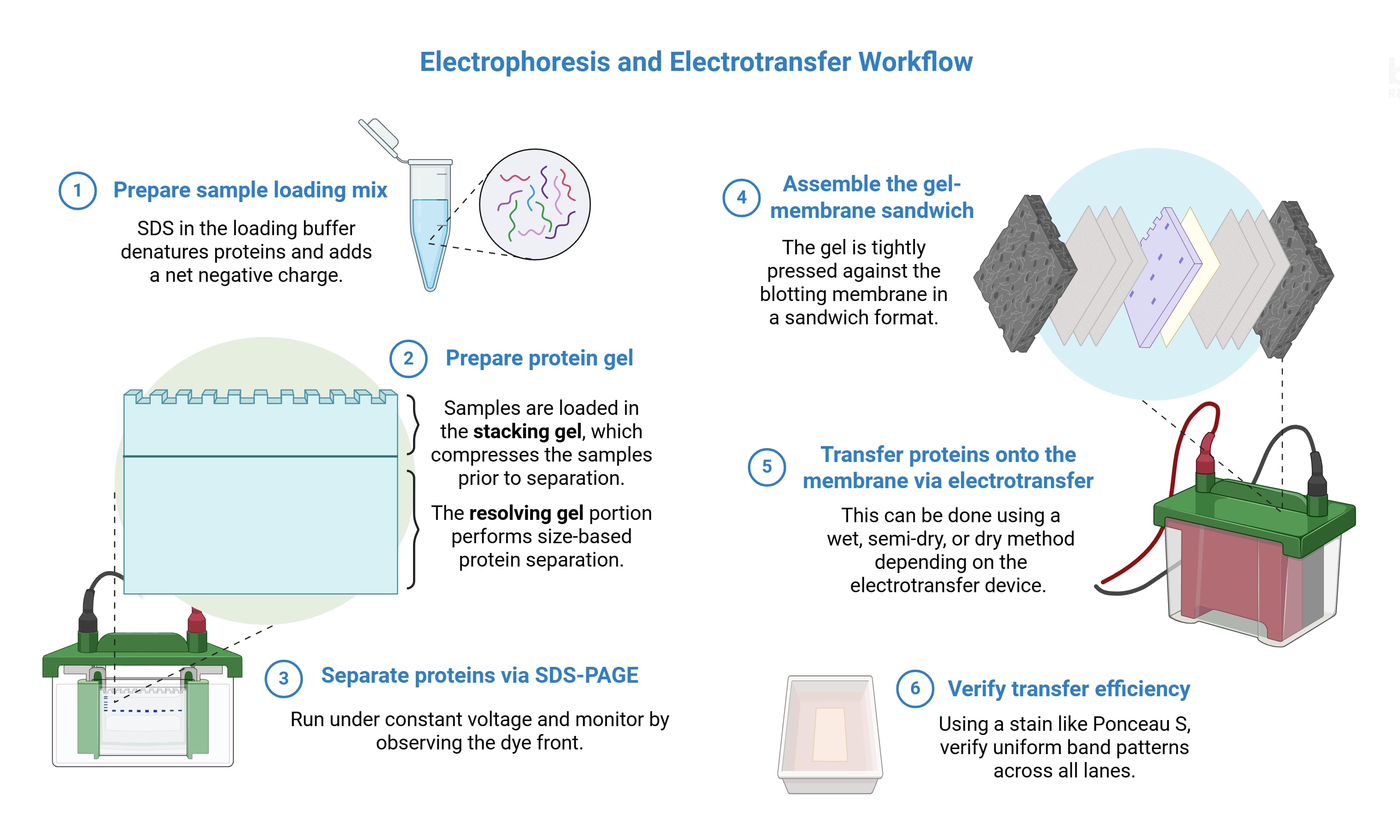
Created in BioRender. Estipona, D. (2026) https://BioRender.com/e73gglq
SDS-PAGE for Western blot
The general protocol for SDS-PAGE can be broadly summarized as follows:
1. Prepare the protein lysate using an ice-cold lysis buffer, or thaw if previously frozen. Quantify the protein concentration in each sample using a validated protein assay, such as the BCA or Bradford assay, to establish the basis for equal loading.
2. Add gel loading buffer to each protein sample. SDS gel loading buffers typically contain a tracking dye, SDS, and a reducing agent such as DTT or 2-mercaptoethanol to disrupt disulfide bonds.
3. Denature the proteins by heating the samples, generally at approximately 95-100°C for 5 to 10 minutes. Samples containing hydrophobic transmembrane proteins may require lower denaturation temperatures to avoid heat-induced aggregation.
4. Cast the polyacrylamide gel if not using a precast gel format. Hand-casting requires careful assembly of casting plates, typically using a dedicated casting device for proper alignment and clamping. Gel formulations generally include deionized water, acrylamide/bisacrylamide solution, SDS, an appropriate buffer (commonly Tris-HCl), and the polymerization initiators APS and TEMED, which are added last.
SDS-PAGE employs a discontinuous buffer system and is prepared in two distinct portions. The resolving gel, which carries out the separation, is cast first and can be formulated at a fixed acrylamide percentage (commonly 10%) or as a gradient (often 4-20%). The stacking gel is poured on top and contains a lower acrylamide concentration, typically around 5%. This upper portion forms the loading wells and functions to compress samples into tight, discrete bands before they enter the resolving gel, improving resolution.
5. Assemble the gel into the electrophoresis device and fill the chambers with an appropriate running buffer such as Tris-glycine-SDS, MOPS, or MES, which maintains pH and conducts current during the run.
6. Load equal amounts of protein per lane, typically between 10 and 40 μg, by placing the pipette tip just above the bottom of each well for precise delivery. Include a prestained molecular weight marker in a dedicated lane to allow visual tracking of protein migration during both electrophoresis and membrane transfer.
7. Run the electrophoresis system under constant voltage, commonly starting at 80 V through the stacking gel and increasing to 120-200 V through the resolving gel. Proteins will migrate toward the positive electrode at the bottom of the gel. Monitor the run closely and stop once the dye front approaches the bottom of the gel to avoid loss of small proteins.
Western blot transfer overview
Following electrophoresis, the transfer step moves proteins from the polyacrylamide gel onto a solid blotting membrane, making them accessible for antibody-based detection. This process relies on an electric field oriented perpendicular to the gel surface, which drives the negatively charged, SDS-coated proteins out of the gel matrix and onto the membrane. In practice, this is achieved by assembling the gel and membrane into a sandwich and applying a current using an electrotransfer device. Traditionally, this has been carried out using the wet transfer method, in which the entire assembly is fully submerged in transfer buffer. Notably, semi-dry and dry transfer systems, which often use proprietary concentrated buffers, have seen growing adoption in recent years due to their ability to achieve efficient transfer in significantly reduced times.
Transfer efficiency is highly dependent on the molecular weight of the target protein. Large proteins (>150 kDa) generally require extended transfer times or higher current, and benefit from the use of lower-percentage gels to facilitate migration out of the matrix. Conversely, small proteins are susceptible to over-transfer, passing through the membrane entirely if the transfer time or voltage is excessive.
Western blot transfer protocol
The protocol for Western blot transfer generally includes the following key steps.
1. Prepare the transfer buffer, which is typically formulated with Tris and glycine to maintain pH and ionic conductivity. Methanol is often included at up to 20% to limit gel swelling and improve protein binding to the membrane, though its concentration may need to be reduced when transferring high-molecular-weight proteins.
2. Prepare the blotting membrane by equilibrating it in transfer buffer. If using PVDF, the membrane must first be pre-wetted in methanol or ethanol before equilibration, as PVDF requires activation prior to use.
3. Assemble the gel and membrane into a sandwich by pressing them firmly together, layering between pre-soaked filter papers and sponges. All air bubbles must be removed using a roller, as bubbles will prevent local protein transfer and result in blank regions on the membrane.
4. Load the sandwich into the electrotransfer device with the correct orientation: the gel facing the cathode and the membrane facing the anode, so that negatively charged proteins migrate toward the membrane. From this point forward, the membrane must be kept wet at all times and must not be allowed to dry prior to signal detection, as drying can irreversibly alter antibody binding.
5. Apply current to initiate the transfer. Run duration is typically around one hour but varies depending on the transfer system and the molecular weight range of the target proteins. Samples containing high-molecular-weight or hydrophobic membrane proteins may require extended transfer times or modified conditions.
6. After transfer, verify efficiency by staining the membrane with a reversible total protein stain such as Ponceau S. Uniform band patterns across all lanes confirm adequate and consistent transfer. Ponceau S can be removed by washing with distilled water or TBS prior to proceeding with immunodetection.
7. The membrane can be used immediately for immunostaining or stored short-term in buffer at 4°C. Storage should not exceed one week, as prolonged storage risks protein degradation or diminished signal.
PVDF vs nitrocellulose membranes
Polyvinylidene fluoride (PVDF) membranes are characterized by their high mechanical strength and durability, which makes them the preferred choice for experiments requiring the stripping and re-probing of a single blot. They typically offer a higher protein-binding capacity than other materials and are particularly effective for detecting low-expressed proteins or hydrophilic antigens. However, because PVDF is naturally hydrophobic, it must be activated with methanol (or ethanol) prior to use to facilitate protein binding. While PVDF is robust enough for advanced applications like amino acid sequencing, it may occasionally produce higher background staining than other options, requiring careful optimization of the blocking and washing steps.
Nitrocellulose membranes are widely used in Western blotting, favored for their high protein-binding affinity and low background signals. Unlike PVDF, nitrocellulose is hydrophilic and does not require methanol activation, allowing it to be pre-wetted simply with distilled water or transfer buffer. Nitrocellulose membranes are often preferred for hydrophobic antigens and are compatible with a wide range of detection methods, including chemiluminescence, chromogenic, and fluorescence. However, nitrocellulose is brittle and prone to tearing, requiring much more careful physical handling. While it provides excellent results for many applications, it is not ideal for protocols that involve multiple rounds of stripping.
References
Taylor SC, Rosselli-Murai LK, Crobeddu B, Plante I. A critical path to producing high quality, reproducible data from quantitative western blot experiments. Sci Rep. 2022;12:17532. doi:10.1038/s41598-022-22294-x.
Pillai-Kastoori L, Schutz-Geschwender AR, Harford JA. A systematic approach to quantitative Western blot analysis. Anal Biochem. 2020;593:113608. doi:10.1016/j.ab.2020.113608.
Ha SG, Park JH, Kim MY, Lee SJV. Brief guide to western blot assays. Molecules and Cells. 2025;100297. doi:10.1016/j.mocell.2025.100297.
Brooks HL, de Castro Brás LE, Brunt KR, et al. Guidelines on antibody use in physiology research. Am J Physiol Renal Physiol. 2024;326:F511-F533. doi:10.1152/ajprenal.00347.2023.
Mishra M, Tiwari S, Gomes AV. Protein purification and analysis: next generation western blotting techniques. Expert Rev Proteomics. 2017;14(11):1037-1053. doi:10.1080/14789450.2017.1388167.
Sule R, Rivera G, Gomes AV. Western blotting (immunoblotting): history, theory, uses, protocol, and problems. Biotechniques. 2023;75(3):99-114. doi:10.2144/btn-2022-0034.
Sormunen A, Koivulehto E, Alitalo K, Saksela K, Laham-Karam N, Ylä-Herttuala S. Comparison of automated and traditional western blotting methods. Methods Protoc. 2023;6(2):43. doi:10.3390/mps6020043.
Mason E. How to improve western blot reproducibility. Biocompare. July 18, 2023. Accessed March 2026. https://www.biocompare.com/Editorial-Articles/597371-How-to-Improve-Western-Blot-Reproducibility/
Li C, Guan J, Li Y, et al. Protocol for high-sensitivity western blot on murine hematopoietic stem cells. STAR Protoc. 2023;4:102578. doi:10.1016/j.xpro.2023.102578.
Tanabe L. The ultimate quick tips & tricks guide to western blots. Biocompare. May 09, 2024. Accessed March 2026. https://www.biocompare.com/Editorial-Articles/612258-The-Ultimate-Quick-Tips-Tricks-Guide-to-Western-Blots/
Mason E. Western blot detection of cell membrane proteins. Biocompare. March 25, 2025. Accessed March 2026. https://www.biocompare.com/Editorial-Articles/618237-Western-Blot-Detection-of-Cell-Membrane-Proteins/
Vero Li J, Zhou Z, Cox CD. Western blotting and immunoprecipitation of native human PIEZO1 channels. Bio-protocol. 2025;15(14):e5385. doi:10.21769/BioProtoc.5385.
Liu D, Wu H, Cui S, Zhao Q. Comprehensive Optimization of Western Blotting. Gels. 2023;9:652. doi:10.3390/gels9080652.
Tanabe L. The next generation of western blot normalization. Biocompare. August 01, 2024. Accessed March 2026. https://www.biocompare.com/Editorial-Articles/615714-The-Next-Generation-of-Western-Blot-Normalization/