What is organ-on-a-chip?
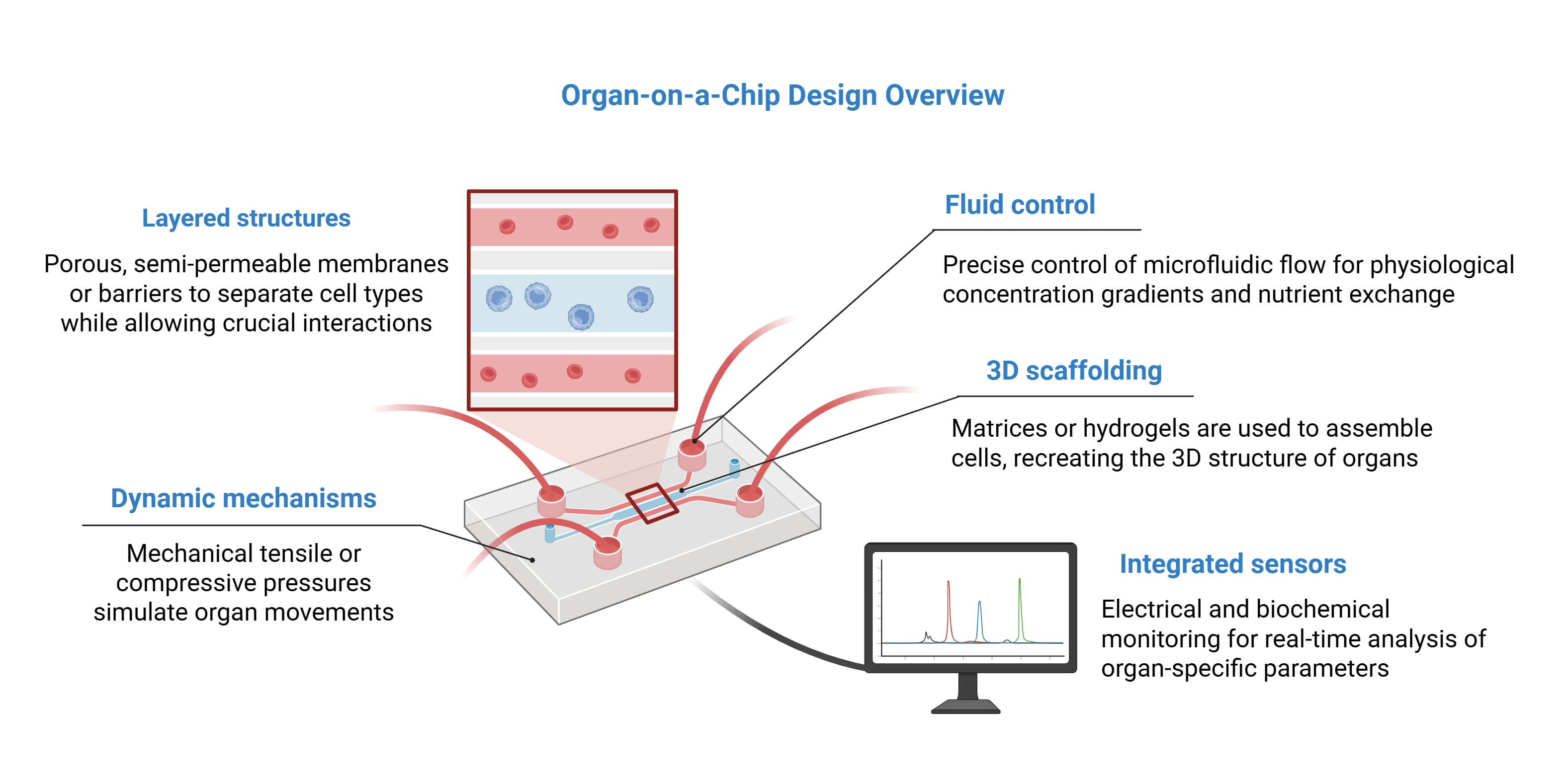
Organ-on-a-chip (OOC) technology utilizes microfluidic platforms to simulate the complex physiological and pathological environments of human organs. As a 3D model, OOC addresses the limitations of traditional 2D cell cultures while retaining the relative simplicity of in vitro culture. Minute fluid volumes are manipulated to maintain stable concentration gradients, reconstruct native cellular microenvironments, and replicate organ functions. Often, this is done by simulating key functional units of an organ, such as the tissue-tissue interfaces between specialized cells. OOCs can also incorporate dynamic mechanical forces, such as fluid shear stress from blood flow and mechanical strain from organ-specific movements, such as lung respiration or intestinal peristalsis. The system can generally execute precise control over necessary culture requirements, such as biochemical gradients, oxygen levels, and nutrient supply, with recent advancements even allowing vascularized tissue structures. Finally, real-time, high-resolution data generation offers rapid analysis of complex biochemical and metabolic activities.
Created in BioRender. Estipona, D. (2026) https://BioRender.com/w2r1bz0
A wide range of human organs and tissues have been replicated using microfluidic technology, including lung, kidney, heart, liver, bladder, pancreas, intestines, brain, as well as tumor tissue. OOC platforms are promising tools to model organ biology, accelerate drug discovery, and improve toxicity assessments. When utilizing patient-derived cells, they can also be used to predict individual therapeutic responses and advance personalized medicine.
Organ-on-a-chip vs. organoids
While organoid and organ-on-a-chip (OOC) models both serve to recreate organ-specific characteristics, OOC platforms offer several unique benefits. Dynamic processes can be readily manipulated with microfluidics, including viscosity, shear stress, fluid dynamics, concentration gradients, and mechanical strain. OOC models can overcome technical limitations of traditional organoids. For example, stomach-on-a-chip models can control luminal flow to improve nutrient delivery and allow direct access to the interior epithelium, which is often inaccessible in standalone organoids. Traditional organoids lack vascularization, and the limited delivery of oxygen and nutrients can constrain organ-specific functionalities. OOC models with engineered fluid mechanics can remedy or enhance perfusion. Finally, integrated sensors enable real-time visualization and quantitative analysis in OOC systems, which are capabilities that are generally disruptive or challenging to implement with traditional organoids.
Conversely, organoid culture is relatively less technically complex, utilizing common tissue culture materials and equipment. OOC models require specialized components such as microfluidic chips, micropumps and valves, integrated sensors, and organ chips, as well as the necessary technical expertise to operate them. In time, emerging commercial producers of OOC platforms may soon offer streamlined workflows and simplified devices, making the technology more accessible to a broader research or clinical audience.
Liver-on-a-chip
Liver-on-a-chip models generally feature continuously perfused microchannels that provide a steady supply of oxygen and nutrients while removing metabolic waste, mimicking blood flow in the liver. Many models aim to recreate the hexagonal shape of the hepatic lobule or imitate the interstitial space between sinusoidal endothelial cells and hepatocytes to mimic the physiological exchange of substances such as drugs or toxins. Devices often utilize a layered structure where different cell types are separated by a porous, semi-permeable membrane or a bionic endothelial barrier. Sensors are integrated to continuously track metabolic biomarkers such as albumin, urea, glucose, and lactate. Cell types housed in a liver chip can often include hepatocytes, liver sinusoidal endothelial cells (LSECs), Kupffer cells, hepatic stellate cells, as well as cell lines like HepG2. Patient-derived stem cells may also be used to create personalized disease models.
Liver-on-a-chip platforms support diverse applications in drug development and disease modeling. These include modeling key hepatic functions, including albumin synthesis and cytochrome P450-mediated metabolism and assessing hepatotoxicity from drug compounds. They can also model pathological states including hepatitis B infection, fibrosis, and alcohol-induced liver injury. Integration into multi-organ platforms further allows prediction of systemic effects, such as hepatic metabolite-induced toxicity in the heart or kidneys.
Lung-on-a-chip
Lung-on-a-chip (LOOC) models are designed to replicate the alveolar-capillary barrier, the primary functional unit of the human lung. These devices generally consist of two main microchannels separated by a thin, flexible, and porous membrane. The upper channel houses human alveolar epithelial cells (mimicking the air-filled alveoli), while the lower channel contains pulmonary capillary endothelial cells (mimicking blood vessels). To mechanically simulate the expansion and contraction of alveoli during breathing, systems utilize side chambers operated by a vacuum or pneumatic actuators. Vacuum-induced stretching of the membranes also allows cells to experience the same motions experienced during breathing. Continuous gas exchange and fluid perfusion are also key functions of LOOC models.
Lung-on-a-chip platforms enable modeling of infectious and inflammatory diseases including SARS-CoV-2 infection, pneumonia, and pulmonary edema. Specialized airway-on-a-chip variants replicate bronchial microenvironments to study chronic obstructive pulmonary disease and assess aerosolized drug delivery and efficacy. These systems also support toxicity assessments, including nanotoxicity screening and evaluation of environmental pollutants such as particulate matter on respiratory function.
Brain-on-a-chip
Brain-on-a-chip (BOC) models aim to replicate the complex architecture, cellular diversity, and functional attributes of the human central nervous system (CNS). Due to the immense complexity of the brain, current models focus on modeling specific brain regions or functional units, such as the blood-brain barrier (BBB) or the neurovascular unit. Devices often utilize multiple parallel microchannels or chambers separated by thin, flexible, and porous membranes. This serves to physically segregate different cell types (such as neurons, astrocytes, pericytes, and endothelial cells) while permitting chemical and paracrine communication. Microfluidic systems enable stable concentration gradients and simulate processes such as blood flow, interstitial fluid movement, BBB permeability, and nutrient exchange. To assess neural functions, sensors such as microelectrode arrays and microtunnels are used to monitor action potentials and synaptic transmission in real-time.
BOC can serve as useful tools for studying pathological mechanisms and testing therapeutics. They have been used in variety of studies, including axonal regeneration after injury, CNS myelination, BBB penetration by drug candidates and nanoparticles. They are promising models for neurodegenerative processes, such as amyloid-β accumulation, α-synuclein spreading, and motor neuron death. Using patient-derived stem cells, BOC platforms can create tailored models to study rare genetic disorders and predict individual responses to treatments.
Heart-on-a-chip
Heart-on-a-chip models integrate human cells, micromachining, and sensors to emulate the complex architecture and physiological functions of the human heart. These devices typically focus on replicating the mechanical and spatial organization of cardiac muscle tissue. Cardiomyocytes in 3D hydrogel matrices self-assemble into functional cardiac microtissues and are patterned in a linear alignment to recreate mature, anisotropic muscle fiber bundles. Flexible membranes and side chambers operated by vacuum or pneumatic actuators are then used to create pressurized compartments. Deforming the membranes compresses the 3D cell construct against posts to generate cyclic pressure, mimicking physiological heart phases. Integrated microelectrode arrays or microtunnels are used to measure action potentials, conduction velocity, and beat frequency in real-time. Some models feature integrated biosensors to monitor contractile force, extracellular field potentials, and environmental parameters such as pH, temperature, and dissolved oxygen.
Heart-on-a-chip platforms serve as promising tools for a variety of clinical and research applications, ranging from testing the side effects of medications to simulating rare genetic conditions. Among these are studies addressing myocardial infraction and arrhythmias, acute hypoxia conditions, drug responses (to compounds such as epinephrine), and drug-induced cardiotoxicity (such as to chemotherapeutic agents). Heart-on-a-chip systems using iPSC-derived cardiomyocytes from patients with genetic mutations are also promising for studying inherited heart conditions.
Human-on-a-chip
Human-on-a-chip, also known as “multi-organs-on-a-chip,” is an ongoing development aimed to simulate broader human physiology by integrating many related single-organ models in a single platform. Theoretically, the various organs and tissues will be interconnected by bionic blood channels, allowing whole-body system interactions. Among the promises of a human physiomimetic model is the accurate representation of key disease elements that can reduce or eliminate the need for animal models.
Currently multi-organ OOC models are still in the early stages of development. As many as seven OOCs (brain, pancreas, liver, lung, heart, gut, and endometrium) have been interconnected in a study on the effects of tolcapone (a Parkinson’s disease therapeutic) in the human CNS, demonstrating the potential of the human-on-a-chip model to emulate human drug responses. However, to enable wider adoption, the concept will need to address several crucial challenges, such as the need for a more universal cell culture medium, practical sample collection, the increasing complexity with the addition of additional organs, and the challenge of controlling the system’s parameters.
Current challenges
Organ-on-a-chip models face several key challenges that currently limit widespread adoption. Obtaining human cells of sufficient quantity, quality, and genetic diversity remains difficult. It is estimated that only 10-20% of purchased primary cells meet quality thresholds for OOC studies, and available cell lines often lack demographic diversity necessary for population-representative results. Maintaining long-term tissue viability for chronic disease modeling remains challenging, and optimizing co-culture media and parameters becomes increasingly complex as cellular diversity expands, especially in multi-organ systems. To minimize inconsistencies in device design and performance and promote cross-laboratory reproducibility, more standardized manufacturing methods, benchmarks, and validation protocols need to be established. Finally, high initial costs (often exceeding $150,000), time-intensive experimental workflows, requirements for highly specialized technical expertise can contribute to uncertainty on whether the added complexity justifies the large investment, especially when comparing to well-established conventional cell-based methods.