What are spheroids?
Spheroids are dense, three-dimensional cellular aggregates ranging from tens of micrometers to several millimeters in diameter, depending on cell type and culture conditions. Unlike monolayer cultures, spheroids retain endogenous extracellular matrix and cell-cell interactions that partially mimic native tissue microenvironments, including nutrient and oxygen gradients, enhanced survival under stress, and more physiologically relevant signaling. Tumor spheroids derived from cancer cells serve as valuable models for studying cancer biology, evaluating anti-cancer therapeutics, and investigating resistance mechanisms. Spheroids of other cell types have also shown promise in regenerative medicine and the development of cell-based therapies.
Spheroids vs organoids
Spheroids and organoids represent distinct classes of 3D culture models that differ in complexity and derivation. Spheroids can be generated from various cell types, most commonly tumor cell lines or, less frequently, tumor tissue. They form through self-aggregation via cell surface interactions when cultured as free-floating suspensions. Comparatively, spheroid culture is simpler and more straightforward, and several methods are available that do not require specialized media or laboratory equipment. 3D cluster formation typically occurs within days, depending on the method. In contrast, organoids are structurally complex entities that arise from stem cell progenitors. They develop into different cell lineages under the influence of specific physical and chemical cues, intended to recapitulate the architecture and function of target organs. Organoid culture is more technically demanding, requiring extracellular matrix components, growth factors, and multi-step protocols that can span several months for full maturation. Because spheroids are biologically simpler than organoids, this simplicity confers practical advantages for cancer research and drug development. Their ease of culture makes them particularly well-suited for high-throughput drug screening applications. Additionally, tumor spheroids can be co-cultured with other cell types to model processes such as cancer cell migration and invasion.
They offer simplified genetic manipulation and high-throughput screening capabilities. Tissue-derived tumor spheres (TDTS) are generated by partial enzymatic dissociation of primary tumors, retaining robust cancer cell-cell interactions while excluding stromal elements. This makes them effective models of non-vascularized tumor regions. Organotypic multicellular spheroids (OMS) are produced from tumor tissue slices cultured ex vivo under non-adherent conditions. Unlike TDTS, OMS preserve the original tumor architecture including stromal components, vasculature, ECM, and native cell-cell interactions. Finally, tumor-derived spheroids (also known as tumorospheres) arise from clonal expansion of isolated cancer stem cells (CSCs) in suspension culture. They serve as enriched models for evaluating stem cell-related characteristics and therapeutic resistance mechanisms in vitro.
Spheroids for cell-based therapies
Due to their partial recapitulation of a 3D in vivo microenvironment, spheroids are emerging as a promising platform for cell-based therapies. Their dense cellular organization and retained cell-secreted ECM helps to facilitate integration with host tissue. Unlike trypsin-dissociated cells which have disrupted ECM and cell-cell connections, spheroids preserve their native architecture, enhancing survival in ischemic environments and upregulate trophic factor secretion. This enables spheroids to mediate tissue repair through complementary mechanisms, such as by incorporation of functionally differentiated cells into regenerating tissue, or by paracrine signaling that recruits host cells and stimulates endogenous repair processes. Spheroids are also tunable, using parameters such as spheroid size, to influence therapeutic function. Smaller spheroids favor proliferation and direct tissue contribution, while larger spheroids develop oxygen gradients that prime cells for harsh in vivo conditions and enhance paracrine signaling. Spheroid-based therapies are under investigation across multiple tissue types and clinical applications. Hepatocyte, cardiomyocyte, pancreatic islet, and mesenchymal stromal cell (MSC) spheroids have demonstrated tissue-specific morphologies and functions relevant to their native counterparts. Current research explores their application in wound healing, congenital defect correction, and regeneration of bone, cartilage, skin, neural, and cardiac tissues, with different spheroid types at varying stages of preclinical and clinical development.
Spheroid culture workflow
While there are several unique protocols for producing and culturing spheroids, common steps can be broadly summarized to comprise the following steps: cell preparation, seeding, generation, maintenance, characterization, and harvesting.
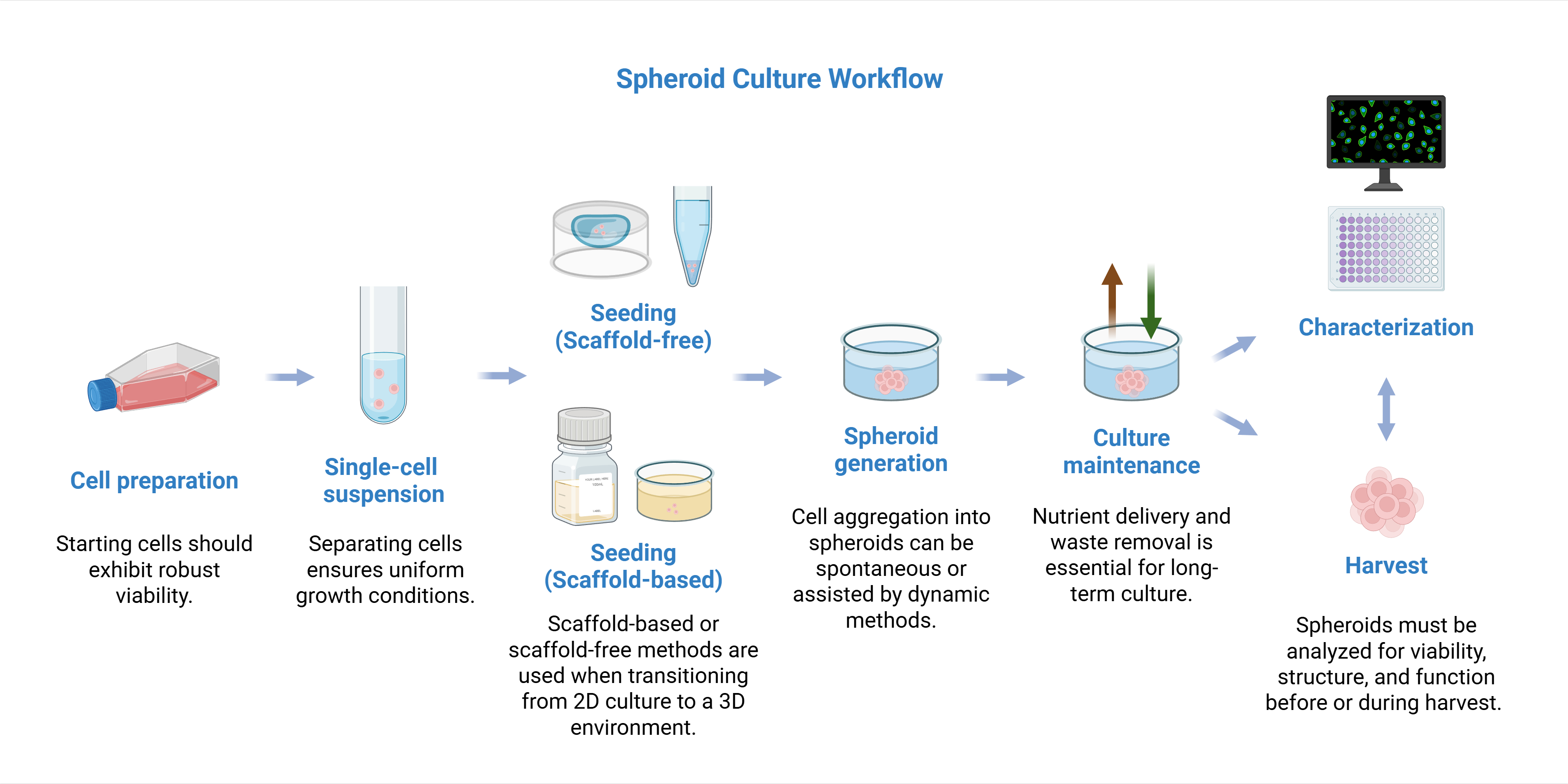
Created in BioRender. Estipona, D. (https://BioRender.com/ma4vszi)
Cell Preparation: Healthy, viable cells should be harvested, then processed into a single-cell suspension to ensure uniform starting conditions. Using a strainer helps to remove cell clumps and debris, which reduces the quality of the starting culture.
Seeding: In this step, cells are introduced to the 3D culture environment. Scaffold-free methods can be used, such as plating in ultra-low attachment (ULA) vessels. More methods are further discussed below. For scaffold- or matrix-based methods, cells are seeded in natural extracellular matrices (ECM) or synthetic hydrogels. Plating densities can vary by cell type and desired spheroid size.
Spheroid Generation: Spheroid formation occurs by spontaneous self-assembly, surface tension, or centrifugation, or other dynamic methods described below. Spheroid formation can occur within 48 to 72 hours, with more challenging cell types requiring more days.
Maintenance: Media should be exchanged within 24 to 48-hour intervals to ensure ample nutrient delivery and waste removal. When using matrices, additional layers of media may need to be periodically added on top of the solidified hydrogel.
Characterization: Spheroids must be assessed for quality and physiological relevance throughout culture, during harvest, or before use in downstream assays. Histology and 3D microscopy, often using confocal imaging with optical sectioning, are used to obtain spatial information. Cellular assays include viability dye staining, apoptosis detection, and cell proliferation assays.
Harvesting: Spheroids are harvested for passage, analysis, or cryopreservation, often using centrifugation and cell strainers. If embedded in a matrix, the culture is mechanically disrupted or dissolved to release the spheroids. Spheroids may also be dissociated back into single cells for passage or quantification.
Scaffold-free spheroid culture
Cells can spontaneously self-aggregate into spheroids in appropriate culture media when adhesion to vessel surfaces is minimized, provided that key environmental factors such as nutrient availability, oxygen tension, and extracellular matrix components are maintained. For cell types that do not readily aggregate, spheroid formation can be induced through various methods that influence structural characteristics, size uniformity, and ultimately, suitability for specific applications. These formation techniques are broadly classified as scaffold-free or scaffold-based approaches. Common scaffold-free spheroid culture methods are highlighted below.
Hanging drop
The hanging-drop method is among the earliest spheroid culture techniques. A small drop of cell suspension at defined density is placed on a culture plate and inverted, where surface tension maintains the drop's shape while gravity drives cells to settle and aggregate at the bottom. Spheroid size can be controlled by adjusting initial cell number, and heterotypic spheroids are readily generated through co-culture. Advantages of this method lie in its simplicity, low-cost, scalability, and ease of imaging. However, its exposed format makes it susceptible to evaporation, requiring labor-intensive media exchange. It can also be challenging to monitor spheroid formation.
Liquid overlay
In the liquid overlay method, cell suspensions are seeded onto low-adhesion surfaces or plates coated with non-adhesive materials such as agarose or polyacrylamide, enabling spontaneous aggregation without cellular anchoring. Specialized vessels, such as those with an ultra-low attachment (ULA) surface, also promotes spheroid self-assembly. Agitation through rocking, shaking, or stirring can enhance spheroid formation by preventing cell settling. In this case, maintaining optimal speed is critical, as insufficient agitation allows spheroids to settle while excessive force damages cells. In addition to the method’s simplicity, its scalability to high-capacity microplates makes it well-suited for large scale screening applications. However, spheroid size, shape, and aggregation kinetics can vary across cell types, and the process offers limited controllability. Additionally, shear forces generated during agitation prevents the use of this method with cells exhibiting low cohesiveness. In microplate formats, these tools offer a simplified method of generating large amounts of uniform spheroids.
Spinner culture
Spinner flasks and rotating bioreactor systems are ideal for large-scale spheroid production. This method utilizes a continuous rotation that prevents cell settling while promoting aggregation. It also allows for consistent oxygen and nutrient delivery along with efficient waste removal, which supports high cell viability and long-term culture. Bioreactors further enhance this approach through continuous medium recirculation, and production capacity scales readily with vessel size. However, the inability to visualize spheroid formation during culture compromises process control, and the resulting spheroids typically exhibit irregular borders and heterogeneous size distributions.
Pellet culture
Pellet culture is a relatively simple approach that utilizes centrifugal force to concentrate cells at the bottom of a centrifuge tube, promoting cell adhesion and aggregation. Cell aggregates can subsequently be transferred to non-adherent coated plates for further culture to complete the spheroid formation. When seeding density is carefully controlled, pellet culture generally produces reproducible spheroid sizes and volumes. Spheroid formation is relatively rapid, requiring only a few minutes of centrifugation followed by a 24-hour incubation period. However, the method is labor-intensive and requires manual precision for spheroid uniformity, while consuming numerous centrifuge tubes that limit scalability. Centrifugal shear forces can damage cells, and spheroids cannot be visualized during growth. Additionally, the typically large spheroid size (>800 μm) may be restrictive for some applications.
Magnetic levitation
Using the magnetic levitation approach, cells are first labeled with magnetic nanoparticles and seeded onto culture plates with low-adhesion surfaces. An external magnetic field then drives cell attraction and aggregate formation, allowing spheroids to form in as little as 24 hours. With higher capacity well-plates, the method is readily scalable for large-scale production. The method also allows for considerable control, enabling the engineering of multicellular constructs with defined shapes and spatial arrangements, with spheroid size directly proportional to initial cell number. However, the introduction of magnetic nanoparticles may pose limitations for sensitive cell types or specific applications, and larger aggregates tend to adopt cylindrical rather than spherical geometries due to non-uniform magnetic field distributions.
Acoustic manipulation
Acoustofluidics, or acoustic levitation, is a more recent approach in spheroid production, in which standing waves are manipulated to trap and organize cells in vessels with protein-repellent amphiphilic coatings. This fast, contactless method enables immediate cell layer formation within seconds and homogeneous spheroid self-organization within a few hours. The resulting spheroids exhibit uniform, reproducible size and shape with enhanced compaction and pore development compared to conventional techniques. It is also capable of mass production, achieving high throughput capacity of up to 12,000 spheroids per chamber. However, the method requires specialized equipment including waveform generators, signal transducers, acoustic reflectors, and acoustofluidic chips, which may limit accessibility.
Microfluidics
Microfluidic devices utilize precisely engineered microstructures to provide physical scaffolds that guide suspended cells into spheroid aggregates. These technologies offer tight control, automation of labor-intensive processes, and high-throughput capacity. Droplet microfluidics, in particular, utilizes cell encapsulation within water-in-oil emulsion droplets with high precision and throughput, generating spheroids of exceptional uniformity. When integrated with automated systems, these platforms can produce thousands of spheroids per hour. They can also be further customized to enhance physiological relevance, such as through designs that mimic systemic drug delivery or facilitate microvascular development. However, the approach requires complex device fabrication, demands greater technical expertise to operate, and complicates spheroid harvesting compared to conventional methods.
Spheroids as cancer models
Tumor spheroids derived from cancer cells have become widely used tools as 3D tumor models. The model serves to recreate key in vivo characteristics including tumor architecture, metabolic gradients, microenvironmental features, and drug response patterns. Compared to conventional 2D cancer models, 3D tumor spheroids have three notable layers: a central necrotic core, an inner quiescent layer, and an outer proliferating layer. This architecture provides greater physiological relevance by modeling oxygen and nutrient gradients, tumor heterogeneity, ECM dynamics, and complex cell-cell interactions that are absent in monolayer systems. The tumor microenvironment recreated within spheroids is particularly significant, enabling more realistic investigations of immunosuppression and therapeutic resistance mechanisms. By bridging the gap between 2D cultures and in vivo models, spheroids have a useful and accessible tool for studying carcinogenesis, tumor progression, and anticancer drug development across many experimental applications. Over many years of tumor spheroid development, several spheroid types have emerged. Multicellular tumor spheroids (MCTS) are monoclonal 3D structures derived from 2D cancer cell lines cultured under non-adherent conditions that promote cell-cell adhesion.
Scaffold-based spheroid culture
While scaffold-free spheroid models have found widespread use due to their simplicity, their ECM architecture remains limited compared to the native in vivo environment. To better recapitulate the complex cell-cell and cell-ECM interactions, hypoxic gradients, and nutrient deprivation conditions found in tumor microenvironments, scaffold-based spheroid models have been developed. Scaffold-based 3D models provide physical support that guides spatially organized cell growth, facilitates ECM deposition, and enables investigation of tumor-stroma interactions. These platforms are particularly well-suited for culturing primary patient-derived cancer cells, conducting drug screening, and assessing therapeutic efficacy on patient samples. Different types of scaffold-based systems are discussed below.
Hydrogels
Hydrogels are three-dimensional networks of hydrophilic polymers that absorb substantial water while maintaining structural integrity. They have become widely used in culturing 3D spheroids due to their ability to mimic extracellular matrix characteristics, such as soft, hydrated environments, tunable mechanical properties, and biocompatibility. Ideal hydrogel scaffolds support cell attachment, migration, nutrient diffusion, and phenotypic modulation with low antigenicity and controlled biodegradability. Hydrogel scaffolds can be derived from natural, synthetic, or hybrid polymer systems. Naturally-derived polymers such as Matrigel, gelatin methacrylate (GelMA), silk, alginate, methylcellulose, chitosan, and gelatin offer inherent biocompatibility that promote native cellular interactions. On the other hand, synthetic polymers such as polyethylene glycol (PEG), polyvinyl alcohol (PVA), and polyvinyl pyrrolidone (PVP) provide improved mechanical strength, stability, and tunability, though they lack intrinsic bioactivity. Extracellular matrix components such as collagen, hyaluronic acid, laminin, fibronectin, and proteoglycans are often incorporated into hydrogels to enhance cell-matrix interactions.
Decellularized scaffolds
Decellularized scaffolds are created by removing cellular components from tissues or organs. This results in a cell-free matrix that still retains tissue-specific ECM composition and architecture. The preservation of native biochemical cues and structural organization creates physiologically relevant environments for generating spheroids for in vitro disease models and for evaluating drug responses. Decellularized scaffolds are a form of patient-derived scaffold that supports personalized therapeutic responses. These platforms have been applied across multiple cancer types to study tumor progression, validate anti-cancer agents, and investigate resistance mechanisms.
Microsphere scaffolds
Microsphere scaffolds provide uniform pore sizes, interconnected architecture, and high surface area, making them versatile platforms for both 3D spheroid culture and drug delivery. In cancer therapeutics, microsphere-loaded scaffolds enable sustained, localized release of chemotherapeutic agents, reducing systemic toxicity while achieving high drug concentrations at tumor sites. As 3D scaffolds, microspheres recapitulate key features of the tumor microenvironment, supporting cell proliferation and cell-cell interactions through their biomimetic architecture. In this platform, pore size and interconnectivity are key variables that govern oxygen and nutrient diffusion throughout the scaffold, directly influencing uniform cell attachment and viability. These properties have positioned microsphere scaffolds as valuable tools for high-throughput drug screening and developing localized therapies for tumor eradication and tissue regeneration.
Nanoparticle Incorporated Scaffolds
The architecture of nanoparticles provides a high surface area-to-volume ratio, offering efficient transport of oxygen, nutrients, and pharmacological compounds. These materials are often combined with existing 3D scaffolds to enhance physical and mechanical properties, which offer promising applications for cancer therapeutics. In studies with osteosarcoma models, several nanoparticle-scaffold systems have been developed, including hybrid hydroxyapatite (HA) and gold nanorod-incorporated hydrogels, HA-enhanced gelatin microribbon scaffolds, photo-cross-linked gelatin methacryloyl (GelMA) hydrogels loaded with gemcitabine hydrochloride-containing liposomes, and nanohydroxyapatite-collagen type I matrices. These platforms demonstrate the versatility of nanoparticle integration in creating biomimetic microenvironments that recapitulate key features of the tumor niche while enabling controlled therapeutic delivery.