Reporter cell lines enable researchers to investigate complex biological processes by producing an easily measurable signal in response to specific stimuli. This article explains how reporter cell lines work, discusses some of the most widely used reporter genes, and highlights some key research applications.
What are reporter cell lines?
Reporter cell lines use precisely controlled gene expression to provide insights into specific biological events. They are typically produced by transfecting a stable mammalian cell line (e.g., Jurkat, THP-1, or CHO) with a plasmid vector in which the reporter gene is positioned downstream of a regulatory element, such as a promoter or a defined protein interaction domain, as shown in Figure 1. In response to specific cues, the regulatory element drives reporter gene expression to produce an enzyme or fluorescent protein that can be easily detected with established laboratory techniques.
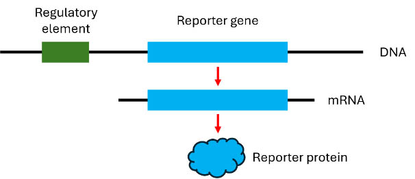
Figure 1. Schematic representation of a reporter gene construct. In response to signaling cues, the regulatory element drives reporter gene expression to produce a measurable readout.
Common reporter genes
One of the most common types of reporter genes are the luciferases, a class of oxidative enzymes found in fireflies, click beetles, and various marine organisms that controls bioluminescence. By converting luciferin to oxyluciferin in the presence of ATP and oxygen, as shown in Figure 2, luciferases generate light that can be quantified using a luminometer and directly correlated to the level of gene expression. A major advantage of luciferase reporter genes is that the signals they produce are virtually free of unwanted background, owing to the fact that mammalian cell lines do not intrinsically generate luminescence.
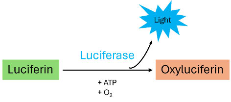
Figure 2. The luciferase reaction
Fluorescent proteins, such as Green Fluorescent Protein (GFP), Red Fluorescent Protein (RFP), and many other more recently discovered variants are also widely used as reporter genes. Following laser excitation, fluorescent proteins emit light at specific wavelengths, which can be detected with either a fluorescence microplate reader or a fluorescence microscope. Fluorescent proteins are especially useful for experiments that require multicolor analysis. However, fluorescent proteins have lower sensitivity than luciferases and their performance can be compromised by background autofluorescence and photobleaching.
Other classic reporter genes include the enzymes β-galactosidase, β-lactamase, and Secreted Embryonic Alkaline Phosphatase (SEAP). β-lactamase activity is commonly measured using the Förster resonance energy transfer (FRET)-based substrates CCF2-AM and CCF4-AM, whereby β-lactamase separates the donor from the acceptor, disrupting FRET and producing a fluorescence shift from 520 nm to 447 nm. β-galactosidase expression is monitored through its action on X-gal, a derivative of lactose, which yields a reaction product with a characteristic blue color. SEAP activity is measured using a variety of substrates, including products that generate colored, fluorescent, and chemiluminescent readouts. Because SEAP is detected in the culture supernatant, it is often used for long-term monitoring of gene expression.
In recent years, it has become increasingly common for two different reporters to be combined in the same cell line. A popular approach involves using both luciferase and GFP to generate quantitative and qualitative data, respectively, from the same experiment. In this scenario, GFP functions as an assay control to normalize luciferase activity, improving the accuracy and reproducibility of experimental results.
Key research applications
Reporter cell lines are used extensively in drug discovery and development, where they allow for determining how different compounds influence cellular signaling pathways. Suppose, for example, you were developing a drug to inhibit interleukin-15 (IL-15) signaling, which is dysregulated in the pathogenesis of various autoimmune and chronic inflammatory diseases. IL-15 exerts its effects through binding to the IL-15 receptor and triggering JAK/STAT signaling. This leads to the formation of STAT5 dimers, which translocate to the nucleus and induce the transcription of specific genes by binding to STAT5 response elements. To investigate the effect of your compounds on this process, you might select a cell line that constitutively expresses the IL-15 receptor and transfect it with a vector construct containing a luciferase reporter gene under the control of STAT5 response elements.
Search Cell-line development services Search Now Search our directory to find cell line development services.
Other types of signaling pathways that can be studied using reporter cell lines include kinase signaling pathways, growth factor signaling pathways, nuclear receptor signaling pathways, and G protein-coupled receptor (GPCR) signaling pathways. Reporter cell lines also enable the study of gene expression in real-time. By using the reporter as a surrogate for a gene of interest, researchers can better understand how the expression of that gene is controlled.
Additionally, reporter cell lines are used for antibody-dependent cellular cytotoxicity (ADCC) and antibody-dependent cellular phagocytosis (ADCP) assays, which allow for assessing Fc-mediated effector functions of antibodies in vitro. Many antibody-based therapeutics work by inducing ADCC or ADCP in patients and, by using an ADCC/ADCP reporter cell line in combination with an appropriate target cell line, it is possible to conveniently identify and prioritize potential antibody candidates for further development.
Troubleshooting reporter cell line assays
While reporter cell line assays vary depending on the model system being used, they share several common challenges and potential solutions. Low signal can result from poor quality DNA, which is often resolved by switching from a regular miniprep kit to a transfection-quality miniprep kit for plasmid purification. Failure to optimize the amounts of DNA and transfection reagent is another cause of low signal. On the flip side, signal saturation can be due to using too much DNA or selecting too strong a promoter for your chosen application. Experimental variability is another recurring problem. Ways of addressing variability include making a master mix for your transfection reactions, identifying the correct plate type for your intended readout (e.g., using white plates for luminescence assays and black plates for fluorescence assays), and repeating your experiment several times to ensure you obtain biologically relevant data.
Conclusion
Reporter cell lines are highly versatile tools with utility for studying development, homeostasis, immunity, and disease pathogenesis. When designing your experiment, the origin of the cell line, type of reporter, and intended application should all be taken into consideration.