Discovering an approved drug that effectively prevents, mitigates, or cures a particular disease can be a protracted process that requires multiple screenings of tens of thousands of compounds prior to clinical trials. Despite rigorous assessment, the majority of these potential drugs, around 80%, fail clinical trials. Effective cancer drugs are even harder to come by, with an estimated 95% or higher of compounds unsuccessful at the clinical trial phase.
Why such high failure rates? Sometimes drugs are unsafe for humans, but most of the time, they are simply ineffective. It has become clear that many cell- and animal-based models are not predictive of clinical efficacy, especially when dealing with heterogeneous diseases, such as cancers. There are multiple parts to a drug screening system. There is the compound or drug, the model in which it is being tested, and the readout.
Scientists are increasingly reexamining their model systems. Two-dimensional cell culture is relatively easy and cheap, yet there is growing recognition that this model may share more differences than similarities with the particular tissue and disease it is trying to mimic. Henry Li, CSO at Crown Bioscience, explains how two-dimensional cultures formed our basis of understanding cancer, yet are not predictive of drug response. “[These cells are] adapted to grow on plastic in a monolayer and in the absence of physiological cell-cell interactions that typically happen in 3D and which are known to influence cell signaling, viability, and drug response.”

Image: Elements of an organoid-based drug discovery and development platform needed to successfully integrate these advanced models into oncology programs. Image courtesy of Crown Bioscience.
Using spheroids, or the more defined organoid, potentially sidesteps, or at least reduces, the number of animals required, and conceivably offers made-to-measure medicine for patients. Drug screening using these three-dimensional cellular models is advancing and carving out new therapeutic paths for diseases from cancer to cystic fibrosis and even COVID-19.
Mini organs and the big picture
“Organoids are self-organizing clusters of cells that grow in three dimensions and exhibit architecture and function that can resemble what is seen in tissues and organs,” explains James M. Clinton, senior scientist at ATCC. Li refers to the organoids as “nearly native and functional mini organs in a dish.” Organoids are derived from either embryonic or adult stem cells, which are capable of differentiation into multiple cell types depending on the culture conditions.

Image: Day 30+ organoids exhibit complex morphology, functional secretory cells, and various gastric and intestinal tissue relevant marker expression. In this micrograph secretory cells line the lumen of an intestinal organoid and express MUC1 (green) and E-cadherin, red. Nuclei have been counterstained with DAPI (blue). Image courtesy of ATCC.
“[Organoids] are also genomically stable when kept in culture, therefore ensuring a more physiologically relevant system, which has been shown to predict with high fidelity, patient response in the clinic,” says Li. Even after cryopreservation, this stability persists, “thanks to specific culturing conditions that prevent stem cell exhaustion and promote their self-renewal and differentiation.” And Nick Asbrock, product manager, stem and 3D cell culture, at MilliporeSigma, points out that organoids can also be genetically manipulated using CRISPR technology.
Judi Wardell-Swanson, senior application scientist, InSphero, highlights that organoids are a very specific type of three-dimensional structure. Spheroids are more inclusive and can also be generated from other cell types, such as tumor cell lines and primary cells, with or without scaffolding. “Our preference is to use primary cells, but we also form spheroids from cell-line and PDX [patient derived xenograft] material for oncology models.”

Image: Different types of InSphero 3D InSight™ Microtissues (spheroids) for drug screening are shown here. Each spheroid model is produced from relevant cell types required to replicate the healthy and diseased organ function. Liver cells for NAFLD and NASH, pancreatic islets for diabetes, tumor cell lines, PDX material, and immune cells for Oncology, and liver cells for liver toxicology.
All of the experts underlined the importance of generating organoids (or spheroids) from patient tissues and biopsies allowing for personally tailored medicine. “Organoids hold great promise for toxicological drug and compound screening and one day could reduce the use of non-human animals for the same purpose,” says Clinton. And Li points out that reproducibility is another advantage of organoids. “Biobanks can be generated for repeat study. Additionally, organoids have a lower interplate variation when compared to other in vitro systems, therefore offering higher quality data.”
Better models of disease, more predictive screens
Organoids and spheroids derived from biopsies offer an in vitro model that researchers believe better recapitulates tumor properties, contains the unique genetic signature of specific tumor types, and experiences an environment similar to that of the tumor, including oxygen and nutrient gradients. “Existing established cancer cell lines may exhibit only a narrow subset of these mutations,” says Clinton. The end result is that “instead of being exposed to random drugs, [the patient] can be treated with those that are more responsive to their tumor type,” says Elizabeth Abraham, senior product manager, Corning Life Sciences. Adds Asbrock, “Recently, tumor organoids have been shown to predict how well patients respond to chemotherapies to aid in personalized medicine.”
Tumor spheroids are another option for scientists. Wardell-Swanson says that some organoid models can take weeks to develop and be very heterogeneous by the end of the process. In addition, it can sometimes be challenging to gain access to fresh tissue from hospitals. There can also be ethical issues related to the use of embryonic stem cells (ESCs) for organoids, as well as restrictions and regulations that vary by country. “Each model has its pros and cons. When deciding which is right … consider requirements for throughput, robustness, maturation time and degree of maturation, multi-cell-type interactions, intra and inter batch reproducibility, and compatibility with downstream analysis.”
Regardless, the set-up for spheroid and organoid drug screens is similar to that for immortalized cell lines. Organoid screens will require Matrigel or some other extracellular matrix (ECM) for scale up and in the screen itself, says Hilary Sherman, senior applications specialist, Corning Life Sciences. And assay optimization may be more challenging as organoids can be variable in shape and size. “Data collection and analysis needs to be optimized, especially when using imaging as the endpoint assay read. 3D imaging results in larger datasets that will need specialized manipulation compared to 2D cultures. Even when using luminescence as the readout, scientists need to ensure that the reagent is able to permeate through these 3D structures,” says Abraham.
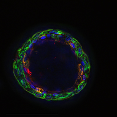
Image: Airway organoid fixed, stained, sectioned, and imaged showing green basal cells, red ciliated cells, orange mucus producing cells, and blue nuclei. Image courtesy of Corning Life Sciences.
Drug screening with organoids typically involves lower density plates compared to 2D screens; for example, rarely will a 1536 well plate be used. Corning offers Matrigel matrix 3D plates as a ready-to-use option for high throughput, as well as a Matrigel formulation designed for use with organoids. Wardell-Swanson says that when 3D models are in suspension, culture vessels and automated liquid handling instruments may require modification to prevent loss of the cell cluster. Infrastructure upgrades may be required. Asbrock says that while there are many bottlenecks for effective drug screening using organoids, “performing some prescreening size and shape steps will help reduce the variability in screening assays.” But the payoff can be worth it in the end. “Several independent studies demonstrated that human 3D models exhibit more clinically relevant responses to drug treatments.”
According to Clinton, ATCC is currently working with the National Cancer Institute (NCI) and the Human Cancer Model Initiative (HCMI) to manufacture and distribute hundreds of novel patient-derived cancer models, including many organoids. These models are molecularly and clinically annotated with data made available by NCI via the Genomic Data Portal and HCMI Searchable Catalog. This can include information such as patient demographics, treatment history, genomic sequences, and mutation status. “At ATCC we are providing detailed culture protocols, media formulations, and other supporting reagents.” They also offer an organoids guide as well as a webinar about how to grow organoids on the site.
CrownBio offers another source of organoid models. Hans Clevers' lab pioneered the technology that allows the derivation of organoids from adult stem cells, and Hubrecht Organoid Technology (HUB) refined it, says Li. “It is currently the only technology available to derive organoids from cancer patients.” CrownBio has an exclusivity agreement with HUB that allows access to its biobank of patient-derived organoids to provide oncology discovery and development services.
Beyond cancer
Organoid usage in drug discovery isn’t limited to just cancer. “Notable other [diseases] are cystic fibrosis, polycystic kidney disease, microcephaly, and infectious diseases of the gut and airway,” says Sherman. And Asbrock notes that COVID-19 has caused increased interest in human lung organoid models. “MilliporeSigma has developed an optimized differentiation protocol along with lung progenitor cells derived from human iPS cells to aid in this research.”
Of course what can be studied in organoid culture may depend on how difficult it is to cultivate certain cell types. For example, all experts noted that intestinal organoids are well established. These can be used for drug absorption and cytotoxicity studies. Asbrock says that a forskolin-induced swelling assay using these organoids can also be used as a phenotypic screening tool for CFTR-Mediated Cystic Fibrosis. MilliporeSigma also has a comprehensive biobank of human adult tissue derived from gastrointestinal organoids for disease modeling created by scientists at the University of Michigan Center for Gastrointestinal Research. “Frozen organoids help new organoid customers produce faster results with less variability than starting from scratch.” The company also recently developed a cell viability assay that works with 3D cultures that offers optimized serum free media for iPSC (inducible pluripotent stem cell) derived organoid. The most up to date list of organoids offered can be found in their site.
Before constructing a screen, scientists must consider the source of the cells, whether adult stem cell, ESC, or pluripotent (PSC). “If multiple sources are possible, consider what the goal is,” says Sherman. “If it is patient treatment, patient-derived adult stem cells may be easiest. If it is to better understand how a disease develops or to compare the disease organoid to the healthy organoid from the same patient, then it may make more sense to start with ESCs or PSCs.” But Abraham says to keep in mind that PSC protocols require additional steps to differentiate into a specific lineage and therefore are more time-consuming and have “higher hit and miss chances to differentiate completely into a specific lineage, which could impact the drug screen.” Asbrock notes, “Organoids derived from adult tissue are thought to contain a more mature phenotype than iPSC-derived organoids and produce more in vivo like responses. However, finding ethically sourced patient tissues can be a problem. Hence, this is why some researchers are interested in using human iPSC-derived organoids.”
Spheroids from primary cells are also getting increased attention in metabolic disease drug testing, says Wardell-Swanson. InSphero uses models derived from human primary cells dissociated from human donor organs. “The major cell types are preserved and recombined in physiologically relevant ratios. The manner in which the tissue aggregates and matures, enables the formation of highly uniform and reproducible spheroidal microtissues with long-lived viability and function.” They currently offer models of non-alcoholic fatty liver disease (NAFLD) and diabetes for drug discovery through partnerships with pharma and biotech companies, as well as a range of toxicology screening services.
Wardell-Swanson has some parting advice. “The rewards of using 3D models for your drug discovery projects will far outweigh the modifications required for implementation of a 3D workflow. Consider any downstream applications and requirements before selecting the 3D model platform and take advantage of existing, established expertise.”
Hero image: Human intestinal organoid culture from patient with cystic fibrosis. Image courtesy of Corning Life Sciences