Reagents, techniques, and technology that support researchers working with primary cell lines are constantly evolving. The result is that more labs are now turning to primary cells, or kindred versions such as hTERT (human telomerase reverse transcriptase) immortalized primary cell lines or iPSCs (induced pluripotent stem cell)-derived primary cells. Today, primary cell culture also looks different. It used to be a feat just to be able to keep these cells alive, even in a static 2D paradigm. But that’s rapidly becoming part of the past. Kevin Grady, senior product line business manager at ATCC, says that scientists who work with primary cells are developing co-cultures of tissue related primary cells and incorporating increasingly complex 3D elements into their culture models. “These model innovations are expected to provide more biological relevance and better mimic the in vivo situation.”
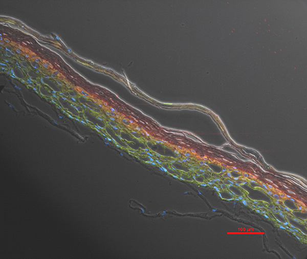
Image: Primary human neonatal dermal keratinocytes seeded on top of Primary human neonatal dermal fibroblasts and induced to form an organotypic epidermal barrier using an air-liquid interface culture system.Image courtesy of ATCC.
Part of the issue with typical 2D cell culture models is that they are too far removed from how cells are aligned and connected within a living organism, notes Katrin Hoeck, associate director, marketing and business development, discovery solutions, at Lonza. The lack of tissue structure and fluid flow leads to altered cell behavior. “As a consequence, static 2D cell cultures are often poor predictors of in vivo cell behavior and its association with disease, drug efficacy, and toxicity outcomes.”
More primary cells available than ever before
Amy Noble, product manager, primary cells & specialty media at MilliporeSigma, explains, “Primary cells are available from more diverse tissue sites than ever, which may make this model more available for a wider range of system/organ/disease studies.” The company offers human primary cells from blood, tumor, and tissue phenotypes.
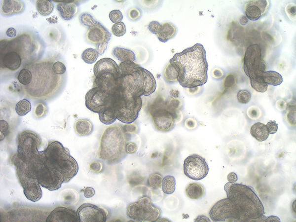
Image: 3D human colon organoid. Image courtesy of MilliporeSigma.
Lonza offers scientists over 150 authenticated human primary cell types from a variety of organ systems and from both healthy and disease donors. And Hoeck notes Lonza has a broad selection of human blood and immune cells that range from peripheral blood mononuclear cells (PBMCs) to isolated T cells or CD34+ cells. She points out that donor variety is critically important “as genes and protein expression often vary across different genetic and epigenetic backgrounds.” As such, Lonza boasts a broad panel of donors. Special donor characteristics, such as human leukocyte antigen (HLA) type, may also be requested. “For diseased cells, detailed donor information including list of medications, cause of death, and more can be requested by contacting our scientific support team.”
Grady says that ATCC also has a large portfolio of human primary cells isolated from both tissue and blood, as well as functionally characterized mesenchymal cells derived from adipose tissue and bone marrow. All of ATCC’s primary cells are cryopreserved at low passage and validated to ensure “high post-thaw viability, extended population doubling level, and high biological functionality.”
Vendors may also offer primary cells from other species. MilliporeSigma, for example, carries primary cells of the murine, feline, and canine varieties.
Customized reagents, technology, and protocols
In addition to more varieties of primary cells available, there are also plenty of customized media, reagents, and kits to make working with these cells considerably easier. ATCC’s Primary Cell Solutions matches basal media and growth factors to specific primary cells to “support and promote cell proliferation and the expression of relevant biomarkers for each cell type.” And Noble says that MilliporeSigma offers primary cells along with “media optimized for maximum relevant growth and longevity of primaries.” When researchers want to use primary cells in 3D systems, there is also their new 3D live/dead assay, which will debut this spring, to help with determining cell health and viability in more complex environments.
Similarly, Lonza also has corresponding culture media for their many primary cells that come in BulletKit™ formats, as well as custom cell isolations, media production, and 3D culture services. Their Nucleofector™ technology allows for non-viral transfection of primary cells at high efficiency with optimized protocols for hundreds of cell types. “A range of Nucleofector devices enable reliable transfection, whether from a single well or when high-throughput screening is required. As the technology allows easy co-transfection of various substrates, it is ideal for genome-editing applications, e.g. with CRISPR, and also allows the use of CRISPR library screenings in primary cells,” says Hoeck.t
Options beyond primary cells still necessary and in demand
Immortalized cell lines still have their place, especially in the early stages of an experiment or when large preliminary screens are needed. “[These] lines are still an important part of cell biology experimental workflow. They are generally easy to culture at a low cost and can be expanded to large numbers,” says Grady. Unfortunately, they are also genetically unstable and tend to have low biological relevance. “As your workflow demands change, experimental variables lessen, and the need for better biological relevance increases, you will likely move on to hTERT [immortalized primary cell lines].”
According to Grady, hTERT lines have the growth characteristics of a continuous line combined with the functionality of primary cells. And since they are isolated from one clone, scientists won’t need to worry about donor variability in their assays. However, Noble notes, “Some researchers wish to avoid introducing exogenous oncogenic proteins into the genome over concerns that it may interfere with cell function or phenotypes. Additionally, genomic drift might be observed in hTERT- immortalized primary cells in culture over time.”
Grady explains that as your experimental design continues to narrow, you might consider moving to iPSC-derived primary cells. “This cell model offers normal diploid genetic stability, primary cell functionality, limited donor variability (derived from a single iPSC clone), but limited ability to expand as these cells have the growth characteristics of a primary cell.”
In addition to providing a larger number of cells compared to most primary cell cultures, iPSCs can be genetically modified to mimic specific disease, according to Hoeck. “They may offer the only true alternative to primary cells, and a more biologically relevant system, in cases when primary cells cannot be obtained by isolation without losing their original functionality. [As is] the case with cardiomyocytes or neurons.”
“As a result, iPSCs are frequently used in target discovery, especially for primary screens, as they offer a more cost-effective model system. For certain toxicity applications, they have also become the model system of choice, the best example being iPSC derived cardiomyocytes, which are commonly used to investigate cardio-toxicity.” But Hoeck says because they do not exactly resemble respective primary cells, validation is needed. She recommends repeating experiments, if possible, in the actual primary cells.
Noble further cautions that while stem cells have many benefits, “they require extensive specialized cell culture training, lengthy differentiation protocols, and may result in immature differentiated phenotypes compared to primary cells.”
Implications for future experimental design
As primary cell availability and support intersect with complex 3D techniques, scientists are creating models and organoids from patient-derived primary tissues. These models have the potential to replace many animal models. According to Hoeck, this includes animals used in testing during drug development. Noble says that MilliporeSigma scientists have developed a 3D skin model using well-characterized keratinocytes and their own media formulation. “We’re particularly proud of this model, as it has significant potential to replace animal models for cosmetics and consumer products, as well as for dermatological research.”
Other innovations on the primary cell front include microfluidic chamber culturing systems. “These chamber systems can accommodate different cell types from the discrete tissues that form a functional organ, leading to organ-on-a chip models. These models could then be ‘connected’ to other microfluidic chambers containing primary cells from different organ systems, leading to the possibility of body-on-a-chip,” says Grady.
However, Grady notes that despite the biological relevance of primary cells, certain in vivo characteristics do get lost when the cells are cultured in vitro. “The next leap would be techniques or technologies that could be used to retain, replenish, or replace these functionalities.”