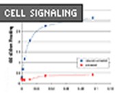
Cell Signaling Technologies’ sandwich ELISA kit to measure phosphorylated histone H3 (Ser10) is a solid phase sandwich ELISA. This ELISA is designed to measure phospho- histone H3 in cell lysates. The ELISA uses a rabbit polyclonal antibody specific for histone H3; the antibody is pre-coated onto a 96-well individual strip microplate. The procedure requires an overnight incubation for best results and consequently, the complete protocol requires approximately 18 hrs. This procedure can be accelerated to 4 hours (without the overnight incubation), but sensitivity is lost. A more detailed description of the procedure follows, but in general: Lysates are added to the microplate and following incubation, the microplate is washed to remove any unbound substances. A rabbit polyclonal biotinylated antibody specific for phosphorylated histone H3 (Ser10) is then added to the microplate wells which binds the captured phosphorylated histone H3 (Ser10) protein. The microplate is washed once again to remove any unbound antibody prior to the addition of HRP-conjugated streptavidin which binds to the biotinylated antibody. At this point, the substrate (TMB) is added to the wells and color development allowed to proceed. Color development occurs in proportion to the amount of phosphorylated histone H3 (Ser10) bound in the initial step. After an incubation period, the color development is stopped and the intensity of the color is measured.
We have used this assay primarily to measure levels of phosphorylated histone H3 (Ser10) in cancer cell lines. The initial step in this procedure is to prepare the cell lysates. Cells are harvested under non-denaturing conditions. The lysis buffer is prepared by diluting the 10X Cell Lysis Buffer (included) and adding 1 mM PMSF. Lysed cells are removed from the plate and transferred to a microcentrifuge tube and kept on ice. The cells are then sonicated for 10 s (also on ice) and then centrifuged at 13000 rpm for 15 min at 4C. The supernatant is then transferred to a fresh tube. The supernatant, which is the lysate, is ready for use in the ELISA or can be stored at -80C for future use. We have substituted the provided lysis buffer with a RIPA buffer with added phosphatase and protease inhibitors cocktails with good results.
Prior to beginning the ELISA, all reagents are brought to room temperature. The first step is to add 100 ul of the provided sample diluent to a microcentrifuge tube, followed by 100 ul of the cell lysate. The microfuge tubes are then vortexed for a few seconds. At this point, 100 ul of the diluted cell lysate is added to the appropriate microplate well. Once all samples are added to the plate, the plate is sealed and incubated for either 2 hrs at 37C or overnight at 4C. We have obtained best results when performing the overnight incubation. After this incubation, the plate is aspirated and washed 4 times with the supplied wash buffer. 100 ul of the detection antibody is added to each well and the plate is incubated for 1hr at 37C. After this incubation, the plate is aspirated and washed once again. The plate undergoes a further incubation, this time with the HRP-conjugated streptavadin for 30 min at 37C. Another wash step is followed by the addition of 100ul of the substrate solution. The plate is then incubated for a further 30 min at room temperature during which time positive wells will become blue in color. The color development is stopped with the addition of the stop solution which turns the blue color to yellow. The plate can then be read in a microplate reader at 450 nm. It is recommended that this is done within 30 min of the addition of the stop solution.
For an ELISA that measures phosphorylation of an intracellular protein and thus, requires cell lysis, this kit is very robust and the intra- and inter assay variability is low. In conclusion, Cell Signaling Technologies’ Pathscan® Phosphor- histone H3 (Ser10) Sandwich ELISA Kit is robust, simple and reliable.
Senior Research Scientist
Discovery Biology
Vitae Pharmaceuticals