Summary
- ELISA (enzyme-linked immunosorbent assay) uses antibodies linked to enzymes to capture and detect a target. An added substrate produces a color that can be measured by light absorbance. First developed in 1971, it remains a low-cost, accessible and highly specific method for protein quantification and biomarker detection.
- Most ELISAs fall into four formats. Direct ELISA is the fastest but least sensitive. Indirect ELISA amplifies the signal with a secondary antibody. Sandwich ELISA captures the antigen between two antibodies for exceptional sensitivity. Competitive ELISA uses an inverse signal and is well suited to small molecules.
- Results depend on several things: antibody affinity and specificity, incubation time, temperature and concentration, microplate material and well shape, the choice of enzyme and substrate, and the reader instrument. Controlling these is key to reproducible data across runs, operators and labs.
First developed in 1971, the ELISA has become a workhorse for detecting analytes of interest within heterogeneous samples in research and clinical laboratories.1,2 Today, ELISA remains a robust, cost-effective, accessible, and highly specific method for protein quantification, and is especially popular for biomarker detection.1 It also serves as an effective bridge between high-throughput screening and more stringent hit confirmation, validation, and characterization. ELISA gives scientists a low-cost method for independently validating candidate biomarkers before committing to more resource-intensive ultrasensitive assays. As with any other laboratory technique, the ELISA method is constantly undergoing adaptation, refinement, and improvement.3
How Does ELISA Work
Generally, ELISA works by capturing antigens of interest with antibodies linked to enzyme-based detection systems.4 Antigen capture is either facilitated by nonspecific adsorption to a suitable surface (typically polystyrene microplates coated with phosphate or carbonate-based buffers) or by targeted binding to an antibody already adsorbed to that surface. The enzyme-linked antibody is then introduced to bind and complex with the captured antigen. Finally, a substrate for the enzyme is added, producing a soluble color corresponding to the amount of antibody present that can be quantified by measuring light absorbance.2,4
Most ELISA protocols fall under one of four categories: direct or indirect ELISAs, which target antigens nonspecifically bound to coated plates, sandwich ELISAs, which capture antigens using target-specific antibodies, and competitive ELISAs, where multiple antibodies are introduced simultaneously and must compete to bind the antigen.2,4
Direct ELISA
In a direct ELISA, the enzyme-linked antibody binds directly to antigen that has been captured and bound to the microplate. Blocking agents such as bovine serum albumin are used to prevent nonspecific binding.4 Direct ELISA protocols involve the fewest steps, making them relatively fast to perform. Furthermore, because direct ELISAs only use one antibody, there is no risk of secondary antibody cross-reactivity. However, direct ELISA offers lower sensitivity than other workflows since there is no antibody-antibody binding-based signal amplification.4
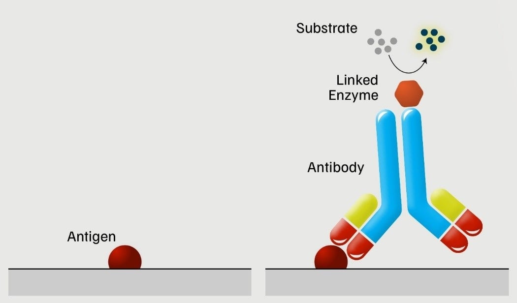
Figure 1: A visual representation of direct ELISA.
Indirect ELISA
Indirect ELISAs are similar to direct ELISAs. However, rather than directly binding the antigen of interest with an enzyme-linked antibody, indirect ELISAs first use a primary antibody to target the antigen and then apply an enzyme-linked secondary antibody to target the primary antibody.2 This allows for signal amplification and greater sensitivity, as multiple secondary antibodies can bind one antigen-primary antibody complex. It also permits more reagent flexibility, as a single secondary antibody can be used across multiple workflows involving different primary antibodies. However, secondary antibody cross-reactivity may negatively affect signal specificity.4
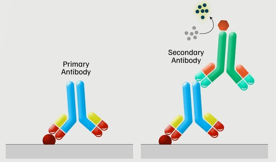
Figure 2: A visual representation of indirect ELISA.
Sandwich ELISA
The sandwich ELISA is named as such because the antigen is captured in between two different antibodies. The first antibody is adsorbed to the microplate during the coating phase so that it can bind the antigen of interest during subsequent sample incubation. The secondary antibody, which is enzyme-conjugated, is introduced after this interaction. Both antibodies specifically target the antigen, which enables exceptional sensitivity but creates additional logistical obstacles: the antibodies cannot target the same epitope.4
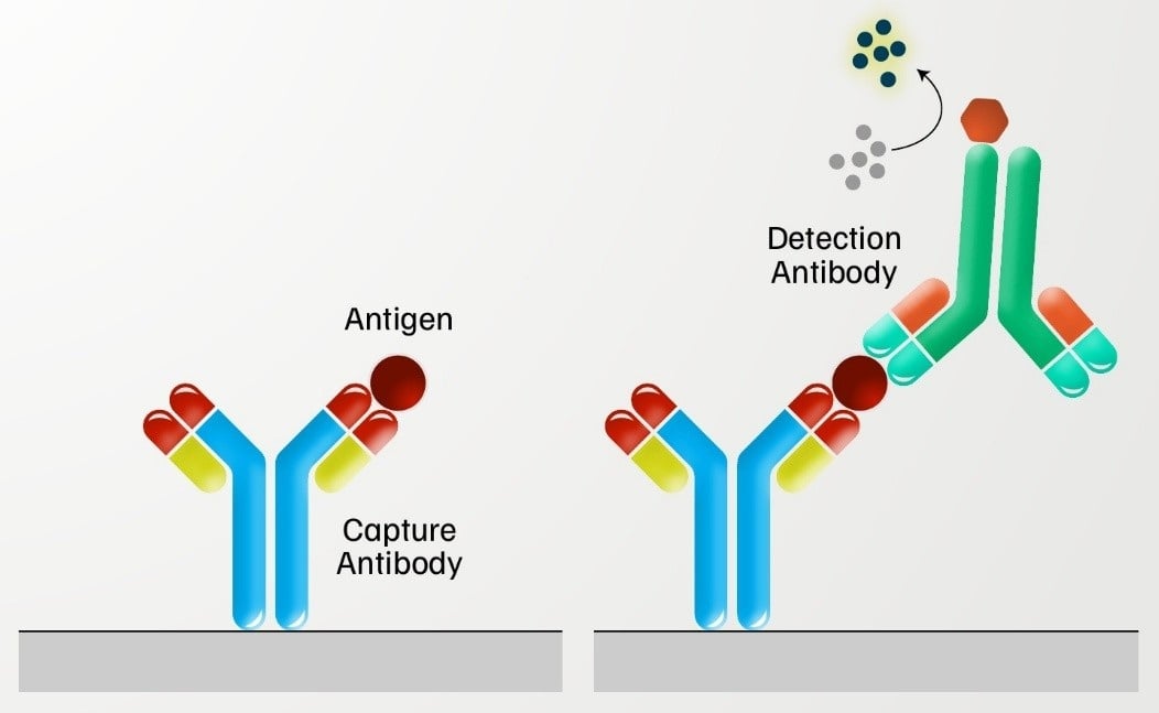
Figure 3: A visual representation of sandwich ELISA.
Competitive ELISA
Unlike other ELISA techniques, competitive ELISA is designed to evaluate antibody presence rather than antigen. Here, the antigen of interest is adsorbed to the microplate and treated simultaneously with the sample and a known amount of antigen-specific enzyme-conjugated antibody.4 A decrease in signal would indicate the presence of antibody in the sample, as it would outcompete the enzyme-conjugated antibody for binding sites. Competitive ELISA is not very sensitive, given its inverse signal output. However, it is ideal for detecting small molecules that are difficult to capture via sandwich ELISA.4
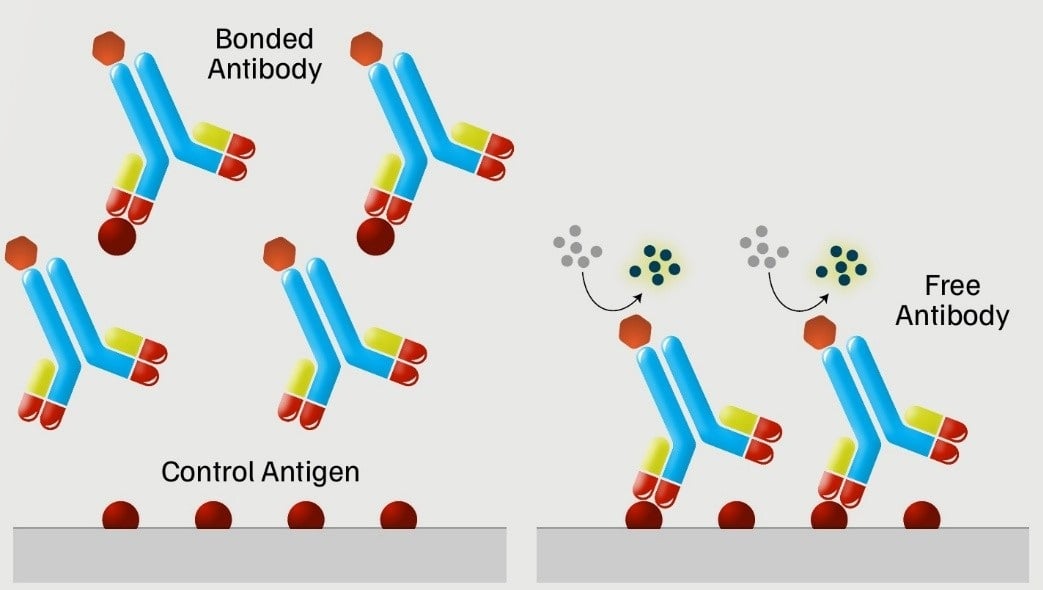
Figure 4: A visual representation of competitive ELISA.
ELISA Troubleshooting
ELISA is well known for its accessibility and general ease of use. However, there are various factors that can affect assay results that scientists need to be aware of as they are planning and performing their assays.2,4 Like all antibody-based techniques, antibody properties such as binding affinity and specificity, as well as incubation parameters such as time, temperature, and concentration, can affect the obtained data. Researchers also need to pay attention to microplate material and well shape, as these can impact coating uniformity. The choice of linked enzyme naturally has a considerable influence on signal generation, as does the amount and reaction efficiency of the added substrate.2 Finally, the data capture instrument—its detectors, filters, and capabilities—plays a major part in data consistency and reproducibility across experimental runs, operators, and research groups.4
References
1. Tighe PJ, et al., “ELISA in the multiplex era: potentials and pitfalls.” Proteomics Clin Appl. 2015;9(3-4):406-22.
2. Aydin S, et al., “An overview of ELISA: a review and update on best laboratory practices for quantifying peptides and proteins in biological fluids.” J Int Med Res. 2025;53(2):3000605251315913.
3. Jeon HB, et al., “Enhancing ELISA sensitivity: From surface engineering to synthetic biology.” Biosensors (Basel). 2025;15(7):434.
4. Zubair M, et al., “Enzyme-Linked Immunosorbent Assay (ELISA) [Updated 2026 Feb 20].” In: StatPearls [Internet]. Treasure Island (FL): StatPearls Publishing; 2026 Jan-.