In theory, genetic disease could be treated by supplying the patient with a correct version of the faulty gene. However, in practice, delivering new genetic material to human cells is difficult. One promising method of gene delivery involves the use of DNA/lipid complexes called lipoplexes. In a study published yesterday in eLife, University of Groningen researchersused advanced simulations to investigate how these lipoplexes deliver DNA fragments into cells. Their results could be used to optimize the method.
Our bodies are very good at destroying foreign DNA, so delivering a new gene to a cell is difficult. One way to do so is by using viruses, but viruses can also trigger the body’s defenses, creating an immune response that can make the patient sick and that can, in rare cases, prove lethal. That is why scientists are now experimenting with lipid-based complexes, which are similar to cell membranes.
Search Antibodies Search Now Use our Antibody Search Tool to find the right antibody for your research. Filter
by Type, Application, Reactivity, Host, Clonality, Conjugate/Tag, and Isotype.
“These lipoplexes are taken up by cells in a structure called an endosome,” explains first author Bart Bruininks. “The problem is that endosomes digest material, so the DNA needs to escape quickly.”
The lipoplex can fuse with the endosome membrane, which then allows the DNA to enter the cell. “This should happen as efficiently as possible to prevent degradation,” Bruininks says. “That is why we want to know exactly how the lipoplex and the endosome membrane interact.”
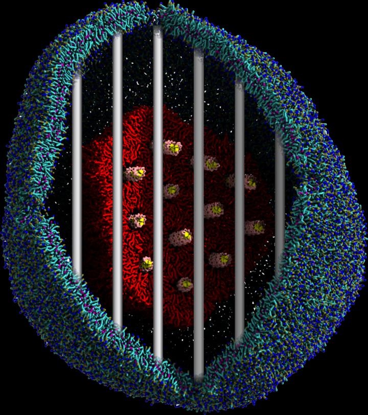
Because this interaction is difficult to explore experimentally, Bruininks and his colleagues created a simulation of the interaction. Using a coarse grain molecular dynamics simulation, they were able to visualize the fusion process between the lipoplex and the endosome membrane as well as the subsequent escape of the DNA.
The lipoplex contains tiny water channels that contain the DNA. The lipids of the lipoplex fuse with the endosome’s membrane. “The lipids form a stalk-like connection and when both layers are fused, a pore opens up that connects the water channels to the cell's interior, allowing the DNA to escape,” Bruininks explains. “This picture of how the process works at the molecular level helps us to understand how to optimize it.”
Image: Visualization of a lipoplex entrapped inside an endosome. The DNA (yellow/pink rods) can only escape upon fusion of the lipoplex (red) with the endosomal membrane (blue).