Modern light microscopic techniques provide extremely detailed insights into organs, but the terabytes of data they produce are often nearly impossible to process. New software called “BigStitcher”—presented today in Nature Methods—can help researchers make sense of these reams of data.
With the help of a few chemical tricks, scientists are now able to render large structures like mouse brains and human organoids transparent. And that’s not all. In 2015, researchers discovered that it was possible to expand ultrathin slices of mouse brains to nearly five times their original volume, thereby allowing samples to be examined in even greater detail.
“With the aid of modern light-sheet microscopes, which are now found in many labs, large samples processed by these methods can be rapidly imaged,” says senior author Stephan Preibisch of the MDC in Berlin. “The problem, however, is that the procedure generates such large quantities of data—several terabytes—that researchers often struggle to sift through and organize the data.”
Search Antibodies Search Now Use our Antibody Search Tool to find the right antibody for your research. Filter
by Type, Application, Reactivity, Host, Clonality, Conjugate/Tag, and Isotype.
To create order in the chaos, the team developed a software program that can be run on any standard computer that somewhat resembles Google Maps. “One can not only get an overview of the big picture, but can also zoom in to specifically examine individual structures at the desired resolution,” Preibisch explains.
BigStitcher can visualize on screen the previously imaged samples in any level of detail desired, but it can also do much more.
“The brighter a particular region of, say, a mouse brain or a human organ is displayed on screen, the higher the validity and reliability of the acquired data,” Preibisch says. And because even the best clearing techniques never achieve 100 percent transparency of the sample, the software lets users rotate and turn the image captured by the microscope in any direction on screen. It is thus possible to view the sample from any angle.
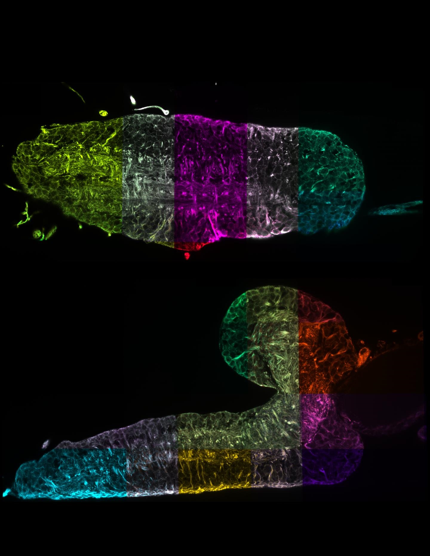
The zoom function allows biologists to find answers to many questions: Where in the brain is cell division currently taking place? Where is RNA expressed? Where do particular neuronal projections end? “In order to find all this out, it is first necessary to get an overview of the entire object of study, but then to be able to zoom in to view the smallest of details in high resolution,” Preibisch explains.
BigStitcher is distributed within the Fiji framework, where any interested scientist can download and use the plug-in free of charge.
Image: Two virtual right-angled cross-sections through the complete nervous system of a fruit fly larva. The combination of expansion microscopy, light-sheet microscopy, and data processing now makes it possible to reconstruct this complex organ optically with nanometer resolution. These data have the potential to track individual nerve cells without complex electron microscopy and thus significantly accelerate studies to investigate neuronal function. Each section highlighted in color is a large 3D image that has been automatically assembled like a mosaic into an overall image that is hundreds of gigabytes in size. Image courtesy of Janelia / MDC.