A collaboration between the University of Wisconsin-Madison and University of Tokyo has led to development of “FluVision,” a means of observing active, live influenza virus infection in the lungs of animals in real time. This work, published today in PNAS, provides a live microscopic view of what happens when a virus infects the lungs and how the body responds in turn.
 "Now we can see inside of the body in real time in virus-infected animals," said study senior co-author Yoshihiro Kawaoka. "It's like we can shrink and go inside the body."
Kawaoka and his team took two strains of flu virus, H5N1 and H1N1, and genetically added fluorescent labels. They called these new fluorescent influenza viruses, Color-flu. They then used two-photon excitation microscopy to image the viruses in vivo. Through this system, the team observed the spread of the virus in the lungs, noting a reduction in blood flow speed in infected areas, as well as activation and behavior of immune cells.
“This approach allowed us to monitor and correlate several parameters and physiological changes including the spread of infection, pulmonary permeability, pulmonary perfusion speed, number of recruited neutrophils in infected lungs, and neutrophil motion in the lungs of live mice,” the team noted.
When mice were infected with H5N1, a highly highly-pathogenic strain also known as “bird flu,” the virus spread more quickly and caused more damage than H1N1, a milder strain. In addition, inflammation-inducing neutrophils were found to increase six-fold when mice were infected with H5N1, compared to the two-fold increase with H1N1.
Interestingly, the team observed that neutrophils displayed two kinds of motion: slow and rapid. In influenza-infected lungs, the neutrophils that remained after the initial infection showed a decrease in rapid motion and spent more time moving slowly, as if scouting for infected cells. This is the first time such behavior has been documented.
"We don't yet know why and what they're doing," said Kawaoka. “We are seeing the mechanisms of the immune system at work. These are the things you discover and it's exciting, but now we have to figure out what's going on."
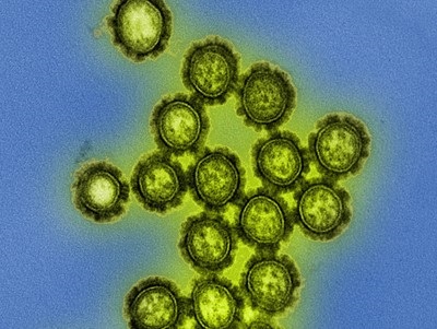
Image: Colorized transmission electron micrograph showing H1N1 influenza virus particles. Image courtesy of the NIH Image Gallery in Flickr.