Multiple sclerosis is an example of a disease caused by myelin degradation. Scientists have questioned how myelin repairs itself and if thinned sheaths are good enough to restore brain circuitry. This week, researchers have published a study that shows in long-lived animals, renewed but thin myelin sheaths are enough to restore the impaired nervous system and can do so for years after the onset of disease. The work can be seen in today's Proceedings of the National Academy of Sciences and comes from the University of Wisconsin, Madison.
Ian Duncan, senior author of the paper, and his team looked at a genetic disorder that affects Weimaraners dogs. This disorder causes 12- to 14-day-old pups to develop tremors and loss of coordination. The disorder is due to the delayed development of the myelin sheath in part of the dog's central nervous system. For most dogs, the symptoms gradually disappear by 3-4 months of age.
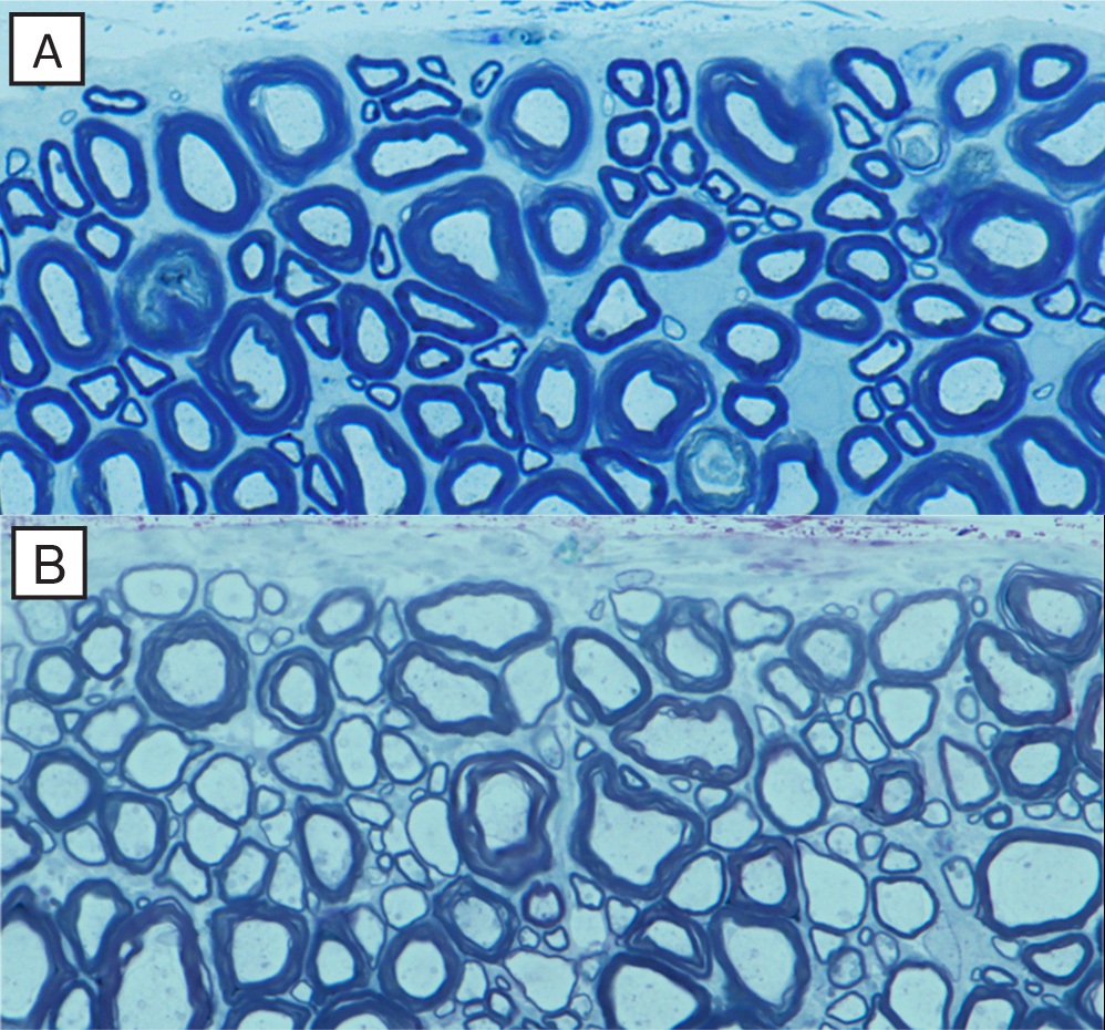
"This is a very widespread mutation in the breed," says Duncan, noting that myelin repair mimicking what is seen in remyelination is known to occur in these dogs as the rejuvenated nerve fibers have a thinned myelin sheath.
Duncan was able to retrieve samples of spinal tissue from two littermate Weimaraner dogs that exhibited these tremors after they lived out their lives. From the samples, Duncan confirmed that thin myelin sheaths persisted and supported normal neurologic function.
 To expand on the data, Duncan looked at optic nerves in cats and was able to show full recovery of the nervous system after demyelination. "We found that nearly every optic nerve fiber was remyelinated with a thin myelin sheath, which is important for understanding human disease because in multiple sclerosis, the optic nerve is often the first to be demyelinated," says Duncan.
The researchers hope that the results will hopefully help with research that is designing therapies that promote myelin repair.
Image: The normal mature dog spinal cord (A) has many axons surrounded by thick myelin sheaths (blue). In contrast, in the recovered dog with the genetic abnormality (B), there are many axons with thin myelin sheaths, identical to that seen in remyelination. Image courtesy of the University of Wisconsin, Madison.