In recent years, three-dimensional (3D) cell models have seen increased uptake for scientific research owing to their greater physiological relevance than traditional 2D cell cultures. However, imaging these complex structures presents technical challenges that are not typically encountered in 2D imaging workflows. We spoke with Karin Boettcher, Product Manager for High-content Screening at Revvity, to learn more about how 3D cell models are being used and pick up tips for capturing high-quality images.
Biocompare: Why have 3D cell models become more popular for scientific research?
Karin Boettcher: "Unlike 2D monolayers, 3D systems better recapitulate in vivo-like microenvironments, including cell-cell and cell-matrix interactions, nutrient and oxygen gradients, and complex tissue architecture. These features result in cellular behaviors that are often more predictive of in vivo physiological responses than those observed in 2D cultures. By better capturing tissue-level organization and function, 3D models help bridge the translational gap between conventional in vitro assays and in vivo studies. This has had a significant impact in areas such as oncology, toxicology, neuroscience, immunology, and precision medicine.
Search Live-Cell Imaging Search Now Search our directory to find the live-cell imaging equipment for your research needs.
"In addition, 3D models enable researchers to monitor dynamic biological processes in real time, such as cell migration, invasion, and treatment response, providing insights into disease progression and therapeutic efficacy that are difficult to gauge from 2D systems. Today, 3D cell models are widely regarded as essential tools across basic research, drug discovery, and toxicity screening workflows, particularly as the industry shifts away from animal testing and toward more predictive, biologically relevant cell models earlier in the research pipeline."
Biocompare: What types of 3D cell models are researchers commonly using?
Karin Boettcher: "Researchers are using a wide range of 3D cellular systems, including spheroids, organoids, and organ-on-chip systems. Each model offers unique capabilities for addressing specific biological questions, and each presents distinct imaging requirements and challenges that influence experimental design and technology selection.
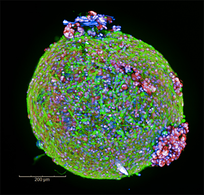
"Spheroids are simple, self-assembled cell aggregates that form sphere-like structures mimicking key aspects of tissue architecture. They are commonly used in cancer research and drug development to evaluate compound penetration, efficacy, and toxicity, offering a relatively simple yet physiologically relevant model. The uniform geometry and controlled size of organoids make them well-suited for high-throughput imaging and automated analysis in screening environments.
"Organoids are more structurally and functionally complex 3D structures that self-organize to replicate key features of specific organs, such as the intestine, liver, brain, or pancreas. They can recapitulate tissue-specific architecture, cellular diversity, and aspects of organ functionality, making them particularly valuable for modeling organ development, investigating disease mechanisms, testing drug responses, and advancing personalized medicine approaches. Due to their inherent morphology and size variability, organoids require advanced imaging techniques to study their complex structure and cellular organization.
"Organ-on-chip (OOC) systems are microfluidic devices containing channels lined with living cells that form miniature organ-like structures. They enable cell-cell and cell-microenvironment interactions that mimic functional units of specific organs, and are often used to study diverse physiological processes under controlled conditions, test drug responses, and gain insights into human biology. The unique architecture of OOCs requires imaging systems capable of accommodating specialized formats and multiple layers within the plates.
"Other 3D structures of interest include co-culture models embedded within hydrogels that incorporate multiple cell types, for example immune cells, fibroblasts, or endothelial cells, to better mimic the tumor microenvironment or tissue complexity."
Biocompare: What are the main challenges for 3D cell imaging?
Karin Boettcher: "One of the primary challenges for 3D cell imaging is achieving high-quality images from large, dense 3D structures. As sample thickness increases, light scattering and absorption become more pronounced, leading to reduced signal intensity, decreased resolution, and loss of image contrast in deeper tissue layers.
"The acquisition of large volumes of image data further increases experimental complexity. Capturing multiple Z-planes and fluorescent channels can significantly extend imaging times, increasing the risk of phototoxicity and photobleaching. Additionally, the generation of large datasets places increased demands on storage capacity, data transfer, and downstream processing capabilities.
"Beyond image acquisition, extracting meaningful insights from 3D datasets requires advanced image analysis approaches. Researchers need to segment and quantify parameters such as volume, morphology, intensity, texture, spatial relationships, and distances in 3D, often across large sample numbers to support statistically robust conclusions.
"Together, these challenges highlight the need for integrated 3D imaging workflows that combine optimized optics, intelligent image acquisition strategies, and efficient data management and image analysis platforms to enable scalable, reproducible, and biologically meaningful 3D cell imaging outcomes.”
Biocompare: Can you share some tips to help researchers capture good 3D cell images?
Karin Boettcher: "Capturing high-quality images from 3D cell models requires a combination of thoughtful sample preparation, appropriate instrument choices, and intelligent image acquisition strategies. Here are six key considerations:
- Choose confocal over widefield microscopy. Confocal approaches remove out-of-focus light, enabling the acquisition of image stacks with improved signal-to-noise ratios and high resolution in all three dimensions (X, Y, and Z).
- Select water immersion objectives. These lenses offer higher numerical apertures than air objectives, so they capture up to four times more light, which improves imaging speed and helps minimize photodamage. They also provide higher resolution in X, Y, and Z dimensions, allowing deeper imaging of 3D structures.
- Optimize your spheroid model size. Spheroids with diameters of ~500 µm or less are generally recommended to ensure intracellular features can be imaged and analyzed.
- Apply optical clearing methods. Clearing techniques can increase the amount of light penetrating 3D models to excite fluorochromes and remove biomaterial that blocks the fluorescent signal reaching the cameras.
- Use multiple cameras to accelerate acquisition. Imaging time can be reduced by using spinning disk confocal imaging systems that combine laser-based excitation with multiple cameras, particularly for 3D cell models that require multiple Z-planes and fluorescent channels.
- Leverage intelligent acquisition tools. Automated systems can pre-scan samples at low magnification to identify regions of interest, then re-scan at higher magnification with each structure centered in the image.
Biocompare: How does Revvity support 3D cell imaging?
Karin Boettcher: "At Revvity, we’ve built a comprehensive portfolio designed specifically for high-content imaging and analysis of 3D cell cultures, helping researchers generate more physiologically relevant data that can drive better-informed decisions.
"Our spinning disk confocal high-content imaging systems, such as the Operetta™ CLS™ and our newest platform, the Opera Phenix OptIQ™, were developed with 3D models in mind. The pinhole distance of our confocal spinning-disk technology is particularly well-suited for imaging spheroids, organoids, and other complex models, enabling the acquisition of image stacks with improved signal-to-noise ratios, high resolution, and minimal sample illumination. Our automated water-immersion objectives also capture up to four times more light than traditional air objectives, allowing researchers to image deeper into 3D structures and capture more detail, faster.
"The Opera Phenix OptIQ high-content screening system elevates 3D imaging performance with high quantum efficiency cameras (>95% QE) for faster multi-color acquisition, advanced laser-based autofocus for fast and robust imaging of organoids and spheroids, and the ability to image multi-layer plates, such as organ-on-chip plates, seamlessly.
"Our software solutions further enhance the imaging and analysis of complex 3D cell models. Harmony™ imaging and analysis software, which drives the Operetta CLS and Opera Phenix OptIQ platforms, provides intuitive tools for exploring cell models in interactive 3D and XYZ viewers, generating Z stack overviews and galleries, producing 3D renderings and movies, analyzing morphologies and volumes in 3D, and calculating z-stack projections (maximum, minimum, and others). The new “Find Organoids” building block within Harmony software allows for the detection of organoids in brightfield stacks with improved object splitting.
"For handling the large datasets generated by 3D imaging, our Image artist™ image analysis and management platform integrates data from all major imaging systems to enable rapid processing, analysis, and sharing of vast volumes of data.
"To complete the workflow, we offer a range of cellular imaging reagents and microplates to support imaging quality. For higher throughput applications, we provide laboratory automation solutions that support upscaling and standardization efforts across different 3D applications."
For Research Use Only. Not for use in diagnostic procedures.