Traditional methods for multiplexed fluorescent imaging of tissue samples present challenges including the following:
- Spectral bleedthrough limits the number of markers that can be detected
- Inadequate sensitivity may result in low-abundance targets being missed
- Autofluorescence can lead to unwanted background signal
- Throughput is restricted by time-consuming, manual staining protocols
- Data analysis is often subjective, with results being based on scores as decided by the human eye
This article looks at some of the tools and technologies that have been developed to overcome these issues, specifically focusing on immunohistochemistry (IHC) applications.
Enhanced sensitivity with Aluora™ Spatial Amplification Kits
Tyramide signal amplification (TSA) is an established method for enhancing the detection of low-abundance targets. It involves incubating the sample with an analyte-specific primary antibody and an HRP-conjugated secondary antibody, followed by the addition of a fluorescently labeled tyramide substrate. HRP catalysis of the substrate causes high-density tyramide labeling at the target site. Because the fluorescent label becomes covalently bound to the sample, multicolor staining can be accomplished by stripping the antibodies away and repeating the process with a different substrate.
Search Cell imaging systems Search Now Search our directory to find the right cell imaging systems for your research needs.
To further increase the sensitivity of TSA, Thermo Fisher Scientific recently launched Aluora™ Spatial Amplification Individual Dyes and Aluora™ Spatial Amplification Kits. “These products let researchers visualize up to eight targets in one tissue sample,” explains Steve Titus, Senior Manager Product Management at Thermo Fisher Scientific. “Importantly, the exceptionally bright Aluora dyes provide up to 200X greater sensitivity compared with traditional IHC and up to 10X greater sensitivity compared with other TSA techniques.”

Cyclic staining using Aluora™ Spatial Amplification Kits. Image provided by Thermo Fisher Scientific.
Easier experimental setup and no photobleaching with Quantum Dots
Quantum dots (QDs) are nanocrystalline semiconductors whose electronic and optical properties are determined by their size and shape. They have several advantages over conventional fluorescent dyes, including superior brightness and much tighter spectral emission, which respectively improve the detection capability and allow for studying more targets in the same sample. “Another key benefit of quantum dots is that multiple probes can be excited with a single laser, which greatly simplifies experimental setup,” reports Kristie Krug, CEO and COO at Core Quantum Technologies.
To support the use of quantum dots in biological applications, Core Quantum Technologies has developed a proprietary technology for encapsulating multiple QDs in a protective polymer coating. This provides resistance to photodegradation and allows for any protein or antibody to be conjugated to the particle surface. “Photobleaching is a common problem for traditional dyes,” comments Krug. “Testing has shown QDs to be stable at room temperature under ambient lighting for two months, with no loss of fluorescence.”
Faster spatial profiling with SignalStar™ Multiplex IHC
SignalStar ™ Multiplex IHC labels up to eight targets in FFPE tissues simultaneously. Briefly, oligo-conjugated antibodies are added in a cocktail to the sample, then complementary oligos with fluorescent dyes are used to amplify the signal of up to four antibodies in the first imaging round. For plex sizes greater than four, the complementary oligos are gently removed (with dsDNase) and the process is repeated to visualize up to four additional oligo-conjugated antibodies. Protocols can be run manually or automated on Leica Biosystems’ BOND RX Autostainer.
“SignalStar Multiplex IHC significantly reduces timelines for panel optimization and validation by using highly optimized protocols and ready-to-go antibodies,” says Sarah Klein, Ph.D., Associate Director, Multiplex Assays, Cell Signaling Technology. “Consequently, researchers can achieve spatial biology data up to 70% faster than traditional multiplex IHC methods. Another major advantage is that SignalStar uses rigorously validated CST® antibodies, which represent the most cited antibodies on the market.”
Simultaneous detection of more markers with Imaging Mass Cytometry™
Imaging Mass Cytometry (IMC™), based on CyTOF® technology, uses antibodies labeled with metal tags for immunostaining samples in a manner analogous to traditional IHC staining. The samples are then introduced into a Hyperion™ Imaging System for data acquisition and analysis. “By replacing fluorophore labels with metal tags, IMC removes the need to worry about spectral overlap or autofluorescence,” says Jennifer Ellis, MS, Director of Scientific Content at Standard BioTools. “As such, 40 or more markers can be imaged simultaneously, for faster time to results compared to fluorescence-based methods.”
To streamline entry to IMC, Standard BioTools has developed a range of ready-to-use IMC™ Panel Kits, featuring a modular design to enable customization for specific research applications. “Our 31 antibody Human Immuno-Oncology IMC Panel is the single largest IMC panel currently available,” reports Ellis. “It is also available in seven modular subpanels to provide more flexible phenotyping options.”
Fully automated spatial multiomics with the MACSima™ Platform
The MACSima™ Platform incorporates MACSima Imaging Cyclic Staining (MICS) technology, an iterative process involving staining, imaging, and signal erasure. Proteins are detected with fluorochrome-conjugated antibodies, and RNA with RNAsky™ Detection Probes, while signal erasure is achieved via photobleaching or controlled fluorochrome release (when using REAdye_lease™ Antibodies, REAlease® Antibodies, or RNAsky Detection Probes). Critically, both signal erasure mechanisms are sample friendly, preserving epitope integrity while efficiently clearing signals for subsequent cycles.
“Researchers can select from over 600 pretested recombinant primary conjugated antibodies, or can integrate their own antibodies for maximum flexibility,” says Dr. Jenia Schlegel, Global Product Manager at Miltenyi Biotec. “We also offer modular panel kits, including ultrahigh-plex, plug-and-play panels for detecting hundreds of protein markers and dozens of RNA markers in the same section. Importantly, the MACSima is fully automated and comes with a user-friendly yet sophisticated data analysis software, which additionally improves experimental reproducibility and minimizes hands-on time.”

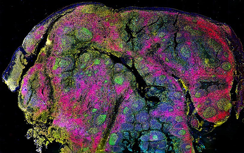
Detection and analysis of 40 protein markers and 27 RNAs on the same non-small cell lung cancer tissue using the MACSima Platform. Image provided by Miltenyi Biotec.
High-resolution detection and unlimited plex with CellScape™
CellScape is an automated platform that performs repeated cycles of staining, imaging, and signal removal via gentle photo-inactivation (with filtered visible light). It streamlines high-plex detection by letting researchers add up to 5 antibodies at a time, to as many as 4 tissue sections, and enables the same fluorophores to be used across multiple cycles. Further time savings can be gained by using pre-validated antibody panels.
“Other ways that CellScape improves on traditional IHC include its high (8-log) dynamic range, which ensures that both abundant and scarce targets are accurately detected in the same sample, and its non-destructive workflow, which allows for future re-interrogation if additional biomarkers are needed,” says Tim Sindelar, Product Marketing Manager at Bruker Spatial Biology. “CellScape also removes the subjectivity from data analysis by using sophisticated algorithms to automatically segment single cells and query the fluorescent intensity of different biomarkers, while retaining the mapped position of each cell within the tissue.” Bruker Spatial Biology recently launched a proprietary reagent, EpicIF, to make the photobleaching method even gentler on tissue, as well as expanded its available antibody portfolio by 10-fold.
CellScape high dynamic range biomarker capture in human tonsil. Image provided by Canopy Biosciences (a Bruker company).