The term protein trafficking describes the shuttling of proteins into and out of cells, and between different subcellular compartments. It is essential to cell function and growth, and its dysregulation has been linked to conditions including cancer, cystic fibrosis, and various neurodegenerative disorders. In today’s climate, the role of protein trafficking in virus assembly and release from infected cells is of huge interest as researchers strive to tackle SARS-CoV-2. In this article, we review the inherent challenges of studying protein trafficking and explore some of the techniques being used for protein trafficking research.
Protein trafficking research presents unique challenges
“Protein trafficking is a constant dynamic process in a living organism, involving numerous interactions. This makes it difficult for researchers to acquire a complete picture of temporal and spatial relationships at any given time,” says Quan Lee, Ph.D., director of R&D at Invent Biotechnologies. Add to that the fact that a lot of those interactions are short-lived, and that many of the structures concerned are incredibly small, and it soon becomes apparent that protein trafficking research can be challenging. Moreover, there is a heavy reliance on over-expression systems for studying protein trafficking, which can often give different results compared to endogenous protein behavior.
Sample types vary considerably
Protein trafficking research can be further complicated by the nature of the material that is chosen for investigation. “A diverse range of sample types are used to study protein trafficking,” explains Patricia Thomson, marketing communications coordinator at StressMarq. “These include both live and fixed cells, cell lysates, tissues, and bodily fluids, with different sample types presenting different challenges. As just one example, GFP-labeled proteins are widely used for live cell imaging but can be hard to interpret due to low signals, high background, and high rates of bleaching.” Imaging live cells can also lead to photodamage, especially if the illumination intensity is too high or the interval between images is too short.
Modern techniques complement established methods
Cell-free systems have long been used to study protein trafficking since they provide researchers with a quick and flexible approach to monitor protein interactions. However, because in vitro findings don’t necessarily translate in vivo, many other methods are employed. Thomson notes that one such technique is split fluorescent protein technology, where distinct proteins of interest are tagged with two fragments of a fluorescent protein (FP); when the target proteins encounter one another, the fragments combine to form a fully functional FP that can be monitored.
“Another exciting system is RUSH, which synchronizes protein transport to allow it to be more easily studied,” says Steph Popa, associate scientist at Abcam. “This promises to advance researchers’ understanding of many different aspects of protein trafficking, including those that currently remain unclear—for example, how cargo is transported within the Golgi apparatus, and how proteins are transported directly from the Golgi to the plasma membrane. Techniques such as RUSH may also help reveal how large proteins such as procollagen are transported.”
Being able to isolate cellular components such as organelles or plasma membranes is also of significant value to protein trafficking research. Traditionally, this has involved a combination of homogenization, density gradient separation, and ultra-centrifugation, making it an extremely laborious process that requires large amounts of starting material and can damage the structures of interest. “A more effective method for cell fractionation is to use spin-column based differential centrifugation and selective precipitation,” reports Lee. “This technology has been cited in multiple journals for its use within protein trafficking research, including a 2018 Science publication describing trafficking of the Nav1.6 sodium channel to the cell membrane in response to chronic pain.”
Advanced imaging technologies support a range of protein trafficking studies
Imaging studies designed to monitor dynamic movements within living cells play a major role within protein trafficking research. According to Scott Olenych, Ph.D., product marketing manager for life science automation and widefield at ZEISS Research Microscopy Solutions, one of the main difficulties lies in selecting a microscopy technique that enables rapid, gentle imaging while also providing the optical resolution needed to detect tiny structures such as endosomes, lysosomes, and virus particles.
Renée Dalrymple, Ph.D., product marketing manager for life sciences laser scanning microscopy, also of ZEISS, notes that advanced imaging techniques typically used for protein trafficking research include laser scanning microscopy (LSM or confocal), spinning disk confocal microscopy (SD), super-resolution-structured illumination microscopy (SR-SIM), single particle tracking (SPT), or lattice lightsheet (LLS). Choosing between these methods depends largely on the goals of the study.
“Laser scanning microscopes are readily accessible in many labs and core facilities,” says Dalrymple. “These are capable not only of imaging structure but can also quantify protein dynamics and interactions using methods such as fluorescence recovery after photobleaching (FRAP) and fluorescence correlation spectroscopy (FCS) to measure diffusion rates, and fluorescence resonance energy transfer (FRET) and fluorescence cross correlation spectroscopy (FCCS) to measure interactions. They are also capable of fast and gentle live imaging, especially when integrated with the Airyscan detector for confocal microscopes. Where resolution beyond ~250 nm is required, SR-SIM is preferred; advances in SR-SIM methodology, such as lattice SIM, have made the technique even faster, gentler, and more applicable to imaging deeper into samples. SPT enables the precise locations of individual fluorescently labeled proteins to be followed as they move within living samples so researchers can learn about trafficking and diffusion rates. SD has historically been one of the most sensitive imaging techniques for fast live imaging; however, for long-term live imaging studies with subcellular resolution, LLS is unmatched in its abilities.”
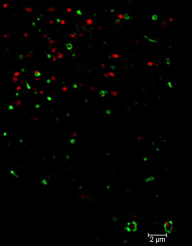
Image: Lattice SIM image acquired with ZEISS Elyra 7 of a U2OS cell expressing an mEmerald-GFP tagged endosomal transport marker (Rab5a) and tdTomato tagged Golgi and Golgi associated transport marker. With this super-resolution technique, it is easy to track the movement of endosomes and the Golgi throughout the cell and to resolve that the endosomal transport marker resides on the membrane.
Novel reagents for protein trafficking research
As researchers learn more about protein trafficking, it is not only detection technologies that have evolved; reagents have also had to keep pace. Abcam has developed a range of knockout-validated antibodies to extracellular vesicle-specific markers including CD9, CD63, and CD81, while StressMarq offers antibodies to VPS35 and DENDD4C—targets related to the retromer, a multimeric protein complex essential for endosome-to-Golgi retrieval of membrane proteins. With knowledge of protein trafficking and its links to disease pathology continuing to grow, the technologies and reagents described here look set to advance further still.