For many researchers, any excitement felt at the prospect of performing an immunohistochemistry (IHC) experiment is rapidly tempered by two key questions: do I really have to carry out so many protocol steps, and how can I simplify the IHC staining process yet still produce data I can trust? Fortunately, help is at hand, with many companies offering expert advice to optimize IHC staining along with practical solutions to streamline protocols and improve the robustness of results. Since one of the main advantages of IHC is its capacity to add spatial context to protein expression, getting it right promises novel insight that can’t be obtained from other immunostaining techniques.
Multiple protocol steps must be considered
“Oftentimes, an immunohistochemistry assay designed to detect a target of interest can be extremely challenging,” notes Yong Zhang, Ph.D., R&D manager at Rockland Immunochemicals. “There are multiple steps in the IHC process that researchers can adapt and modify to improve the quality of their staining, not least the various stages involved in tissue preparation. For example, different fixation and permeabilization conditions should be considered depending on antigen localization, whereas certain tissues may require antigen retrieval to unmask epitopes and allow antibody binding. It may also be necessary to block endogenous targets like biotin or certain enzymes in accordance with the detection method that will be used. On top of all this, identifying a suitable antibody that has been validated with the proper positive and negative controls is critical to the success of IHC.”
Success hinges on antibody selection
When it comes to antibody selection, an error well known to those with a background in technical support and troubleshooting is the mistaken assumption that an antibody that has been validated for IHC with FFPE tissue will work equally well for staining of frozen material. “It is vital that researchers understand there are a number of factors that impact antibody performance, including the means of sample preparation,” explains Katie Crosby, director, immunohistochemistry at Cell Signaling Technology. “Although some antibodies will function appropriately in several applications, others have more limited utility. Functionality in one application neither necessitates nor guarantees performance in another.”
Crosby also points out that while most antibodies will yield some staining in an IHC experiment, it is important that researchers don’t automatically assume this to be specific. “There are certainly still articles being published that describe tissue staining results without also showing that the antibody has been properly validated in that assay,” she explains. “However, with more scientists generally aware of the reproducibility crisis, researchers expect increasingly rigorous in-house validation from antibody manufacturers. At CST, we champion the notion that no single assay is sufficient to demonstrate antibody specificity, and we use multiple complementary strategies to validate our antibodies. These range from binary testing—evaluation of each antibody in biologically relevant positive and negative expression systems—all the way through to supportive strategies such as peptide competition and the use of microarrays.”
Don’t be baffled by blockers
Another common mistake when running an IHC experiment is to use the wrong blocking agent to prevent non-specific antibody binding. To minimize unwanted background staining, it is widely recommended that serum derived from the same species as the host of the secondary antibody is used for blocking and as an antibody diluent, yet this can create confusion if multiple secondary antibodies are employed. “Using a universal blocking agent can eliminate the need to match blocker to antibody species,” says Rajakumar Mandraju, R&D scientist at Enzo Life Sciences. “Moreover, these reagents are often compatible with other applications, providing a level of consistency when comparing data generated by different approaches.”
Minimize background by using labeled primary antibodies
While the nature of IHC has historically dictated that staining protocols are lengthy, one way of reducing the number of steps is to use conjugated primary antibodies. “Directly labeled primary antibodies offer several advantages, provided that the antibodies remain well-characterized and maintain their functionality after conjugation,” says Zhang. “As well as reducing background noise by negating non-specific binding from any secondary antibodies, using labeled primaries also helps to preserve delicate tissues by cutting down wash cycles and sample handling. However, secondary antibodies still have one main advantage: they provide signal amplification to less abundant targets that may otherwise be hard to visualize.”
Nanopolymer detection reagents can boost signal
For researchers struggling to achieve the required sensitivity in IHC, even with secondary antibodies, Enzo Life Sciences offers a range of nanopolymer detection reagents designed to enhance chromogenic detection. “Our POLYVIEW® system is based on secondary antibodies with multiple cross-linked HRP molecules,” notes Mandraju. “This means that the local concentration of active enzyme is several-fold higher compared to non-polymeric HRP conjugates, translating to increased sensitivity. These reagents can be used with the researcher’s choice of chromogen, although we’ve noticed that most IHC experts preferentially use DAB since it provides a strong contrast between signal and counterstain.”
Multiplexed IHC data in just two hours
Of course, another way to get the most out of an IHC experiment is to multiplex it, a concept which Cell IDx has taken to new levels. “Researchers are well aware that the use of fluorescent secondaries is limited by the availability of primary antibodies of different species or isotypes,” notes David Schwartz, Ph.D., Cell IDx co-founder, CEO and CSO. “To address this, we’ve developed a range of modified haptens that we use to label primary antibodies, subsequently allowing for antibody binding to be detected with fluorescently labeled high-affinity anti-hapten secondaries. By combining hapten-labeled primary antibodies in panels of four, tissue alignment of serial sections allows detection of multiple markers via a simple two-hour, two-step staining procedure.”
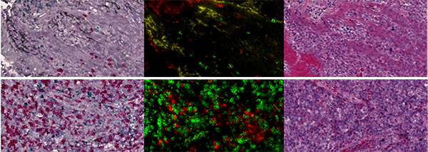
Image: Multiplex chromogenic and multiplex immunofluorescence staining of two tonsil tissues for PD-L1, CD8 and Granzyme B using UltraPlex mxIHC (left: PD-L1 black, CD8 red, GzmB green) and UltraPlex mxIF (center: PD-L1 yellow, CD8 green, GzmB red). Following removal of the coverslip from the mxIF-stained slide, the tissue was H&E stained to allow morphological visualization (right). Image provided by Cell IDx.
Schwartz elaborates that Cell IDx’ modular approach lets researchers mix and match antibodies, all of which share a common staining protocol for ease of use, however he also highlights that continued interest from pharma for chromogenic staining has meant adaptation of the company’s technology to multiplex chromogens. “Like our multiplex immunofluorescence technology, our multiplex chromogenic platform allows development of panels in a primary antibody species-independent manner and has utility to stain serial slides,” he explains. “We’ve recently generated data showing that three chromogens produce similar results to fluorogenic staining; traditional H&E staining can subsequently be employed to increase the information garnered from a single slide. Furthermore, we’ve demonstrated the compatibility of both platforms with the BOND RX autostainer for easy, walk-away operation.”
Careful planning reaps rewards
With a vast range of enabling technologies now available to researchers, IHC is certainly no longer a technique restricted to a handful of experts. Instead, provided due consideration is given to the localization and abundance of the antigenic target, researchers stand a far greater chance of generating meaningful IHC staining data the first time. Whether the experimental aim is to detect a single protein in an isolated tissue sample, or to perform multiplexed staining across a large panel of material, a wealth of tools and advice are out there, ready and waiting to be used.