Immunohistochemistry (IHC) is a technique that uses antibodies to image cells and their components within their appropriate histological context. Over the years, the process has been refined and improved, so various approaches exist that will allow you to acquire a successful IHC image. But just because IHC has been a prevalent laboratory tool since the 1940s doesn’t mean that it is always straightforward.
Common obstacles
“The principal obstacle to good IHC results depends on the level of experience of the researcher,” says Will Howat, head of imaging & immunohistochemistry at Abcam. “For new researchers, or those just starting to work with IHC, it can be difficult to optimize the best protocol from scratch in their lab. Consequently, they may also find it challenging to assess and troubleshoot the technique. To help address this issue, Abcam has created a series of guides to IHC that can help mitigate this.”
Howat adds that for more experienced researchers, the main obstacle to getting good IHC results is finding antibodies that will work on a researcher’s particular application and validating it in the lab. Katie Crosby, director, immunohistochemistry, from Cell Signaling Technology, agrees.
“There are often many antibodies available against a given target, but it is not often the case that each will work perfectly in IHC,” Crosby says. “Complicating matters, most antibodies will yield some staining when applied in an IHC assay. The question is, ‘Is this staining specific?’”
In order to achieve good results, researchers must understand the performance characteristics of their antibodies. Crosby suggests that researchers evaluate data provided by the antibody supplier, considering things like staining on various models, in multiple tissue types, and in negative and positive controls when possible. “It is typically a series of validation assays that together will provide confidence that an antibody is staining specifically,” she notes. “No single assay is sufficient to provide assurance of specificity.”
Crosby also warns that vendor-provided validation data “is a great first step but is not necessarily sufficient.” She says that even excellent antibodies can yield poor results when not handled properly. If the laboratory protocol differs from the vendor’s recommended protocol, Crosby recommends that researchers perform their own validation assays. For more immunohistochemistry tips from Cell Signaling Technology, follow this link.
Detection systems and sensitivity
IHC protocols all depend on a primary antibody that is specific to the tissue of interest; however, there are a variety of detection systems available to create an image. Each detection system has varying degrees of sensitivity.
“A directly conjugated secondary antibody will have the lowest sensitivity, even if conjugated in fluorescence, when compared to a polymer-based detection or multi-enzyme-conjugated-secondary detection,” Howat explains. “Then amplification of a tyramine-based system gives the greatest sensitivity of all, due to the number of detection molecules present in each case. With regards to detection molecules, both alkaline phosphatase and horseradish peroxidase have equal sensitivity, as AP can be cycled for longer. Fluorescence can provide additional sensitivity, but it has added difficulties when applied to highly autofluorescent tissue.”
The type of detection system matters especially when you want to stain more than one epitope in the same tissue. You have to decide between doing a simultaneous stain and a sequential stain, and care must be taken to avoid cross-reactivity between the reagents used.
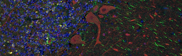
Image: Multiplex immunofluorescence in human cerebellum tissue using recombinant monoclonal antibodies: anti-GFAP (green, ab68428), anti-NeuN (yellow, ab177487) and anti-beta III Tubulin (red, ab52623), DAPI (blue). Image courtesy of Abcam.
When performing a sequential stain, the order of adding detection systems depends on the kits you use. “If these are different (e.g., HRP and AP), the staining system (e.g., DAB or Fast Red) should be employed at the end before the next staining round to minimize the cross-reactivity of different species secondary antibodies,” Howat explains. “If the staining system is the same, additional blocking procedures or stripping steps—such as antigen retrieval to remove the primary antibody complex—need to be employed to minimize cross-reactivity.”
Howat also stresses that controls are important to validate all of these methods in order to ensure that the final image is accurate.
Multiplex staining and fluorescent imaging
 To learn more about multiplex antibody staining and fluorescent imaging, we contacted Stephanie Hennek, R&D manager at Ultivue, a company that specializes in multiplexing immunostaining in situ.
“Multiplex immunofluorescence (mIF) staining is a powerful tool that enables the identification of a wide range of cell phenotypes and mapping of cellular interactions,” Hennek explains. “Within the context of mIF, it is important that fluorescent reporters provide bright, stable signals and do not exhibit bleed-through between channels to allow spectral differentiation of multiple fluorophores.”
According to Hennek, it is common for researchers to spend valuable time and resources “identifying isolated fluorophores and balancing signals between targets.”
Image: Whole-slide image of multiplex immunofluorescence in human non-small cell lung cancer using the UltiMapper I/O PD-L1 kit. Markers detected include CD8 (yellow), CD68 (green), PD-L1 (red), pan-Cytokeratin (cyan), and nuclear counterstain (blue). Image courtesy of Ultivue.
“At Ultivue,” Hennek notes, “we’ve taken the development work out of the researcher’s hands and provide UltiMapper™ multiplex IF assays that are fully optimized and compatible with a wide range of fluorescence microscopes and scanners for whole-slide imaging.”
Hennek provided us with a set of practical tips concerning bleed-through reduction, choosing between simultaneous and sequential staining methods, and mIF controls for successfully carrying out mIF experiments (see box below).
Whether you’re new to IHC or a seasoned user of the technique, you can always benefit from checking your procedure, brushing up your skills, and investigating what new resources and technologies exist. And if you run into trouble, companies like Abcam, Cell Signaling Technology, and Ultivue have support teams available to assist with technical questions regarding any of their products.
Tips to Improve Multiplex Antibody Staining and Fluorescent Imaging
1. Reducing bleed-through between the spectra of fluorophores
Understanding the photophysical properties of fluorophores is key to minimizing bleed-through between channels in a multiplexed experiment. There should be limited overlap between the excitation and emission spectra of the fluorophores that will be imaged together. The filter sets and light sources used in the microscope must be matched to the spectral profiles of each fluorophore in order to isolate the signal from each target.
Oftentimes, spectral unmixing is employed to decipher between bleed-through/crosstalk issues. A consequence of spectral unmixing is that it lengthens the staining-to-analysis workflow and can mask native biology, making it potentially difficult to examine comparative expression levels of specific markers. Ultivue’s kits use spectrally isolated fluorophores to eliminate any chance of bleed-through from the fluorophores.
2. Choosing between simultaneous and sequential staining procedures
From a throughput and biological perspective, it is preferable to stain antibodies simultaneously than to stain sequentially. Sequential staining could potentially block epitope sites depending on the detection system and methodology used. Simultaneous staining allows all antibodies to bind to their epitopes at the same time and reduces the overall assay time. Ultivue’s technology does not require a sequential staining procedure, even for higher plex assays. All targets are stained and amplified simultaneously no matter the number of markers. The simultaneous amplification across all markers allows for the ability to quantitatively assess differential marker expression of the targets in the multiplex panel. Imaging of all targets may be carried out in one or more rounds depending on the number of markers in the multiplex assays with minimal impact on the overall assay-to-analysis time.
3. Important controls for mIF experiments
There are several important controls to ensure a robust and reproducible multiplex immunostaining assay. There should be a control to an established reference standard (e.g., DAB) to ensure fidelity of staining in both monoplex and multiplex formats; positive and negative control tissues to confirm staining specificity for each target; and pre- and post-conjugation staining controls to ensure that the conjugation chemistry did not impact antibody binding efficacy. Ultivue uses all these controls in the assay development process as well as a specially designed TMA with cell lines that have a range of marker expression (e.g., PD-L1), that allows the company to quantitatively assess dynamic range, limit of detection, and limit of quantification.
Tips provided by Stephanie Hennek, R&D manager at Ultivue.