American Peptide Company (A member of the Bachem Group)
Associate Marketing Manager
Alzheimer's disease (AD) is the most common type of dementia and accounts for an estimated 60% to 80% of reported cases. It is estimated that more than 35 million people worldwide have dementia, and this figure is expected to triple by 2050 with the aging global population. Dementia is characterized by a loss or decline in memory and other cognitive abilities. AD is an irreversible, progressive neurodegenerative disorder. A worldwide struggle is underway to find new treatments and therapies to prevent, cure or even slow the progress of Alzheimer's disease. The presence of senile plaques, neurofibrillary tangles, neutrophil threads, beta-amyloid (Aβ) peptide deposits and selective loss of neurons are some of the distinctive characteristics of the disease.
The most common feature of the disease is amyloid plaque deposits. Initially Aβ starts as a solitary molecule and tends to come together in the form of clusters that are still soluble and are capable of free travel in the brain; finally, clusters form the plaques that are the hallmark of AD. Studies show that Aβ has strong binding affinity to the receptors on nerve cells, triggering an intracellular process that erodes their synapses with other nerve cells. Aβ is chemically "sticky" and gradually builds up into plaques. Aβ peptides are proteolytically cleaved from the membrane-bound amyloid precursor proteins (APPs) that are major constituents of these deposits. The peptides result from the APP, which are cut by certain enzymes to yield Aβ. Beta-amyloid molecules can aggregate to form flexible, soluble oligomers that may exist in several forms. It is now believed that certain misfolded oligomers act as seeds to induce other Aβ molecules to take the misfolded oligomeric form, leading to a chain reaction. The seeds and the resulting amyloid plaques are toxic to nerve cells. Plaques and tangles tend to spread through the cortex in a predictable pattern as AD progresses. The familial AD genetic evidence shows a relationship between metabolism of Aβ and AD disease. This progressive brain disorder damages and eventually destroys the brain cells, leading to dementia and decline in other brain functions and eventually leading to the death of brain cells. Alzheimer's is therefore fatal, and unfortunately so far there is no cure.
Characterizing AD
Alzheimer's brain abnormalities, including but not limited to:
- Plaques (microscopic clumps of a protein fragment called beta-amyloid). These sticky brain plaques are known to consist mainly of Aβ and are known to build up in the brain many years before the actual Alzheimer's symptoms start.
- Tangles—twisted microscopic strands of the protein tau. Tangles destroy a vital cell-transport system made of proteins.
- Loss of connections among brain cells responsible for memory, learning and communication. These connections, or synapses, transmit information from cell to cell.
- Inflammation, triggered by the body's immune system.
- Eventual death of brain cells and severe tissue shrinkage, leading to brain lesions.
Beta-amyloid plays a crucial role in AD, and its high concentration (nanomolar to micromolar) in the brain can cause neuronal death. Picomolar concentrations of Aβ slow down the memory and learning processes. The Aβ peptide levels in the brain are dynamically and directly influenced by synaptic activity. Low amounts of Aβ could work as antioxidants because of Aβ’s ability to capture redox metals, such as Cu, Fe and Zn, and thus prevent their participation in redox cycling with other ligands. Aβ peptide has neurotrophic properties and hence many roles, including the growth and survival of neurons, the modulation of synaptic function and defense against oxidative stress. The physiological concentrations of Aβ favor the learning and memory processes.

Figure 1. Amyloidogenic pathway. Image courtesy Cárdenas-Aguayo et. Al [1].
Role of Beta Amyloid in the early detection of Alzheimer's disease:
The role of Aβ40 and Aβ42 peptides in the early pathogenesis of AD has been frequently emphasized in the literature [2]. Aβ40 and Aβ42 are major products of the proteolytic cleavage of the multidomain, integral-membrane, type-1 protein APP, which has important roles in cell adhesion, neuronal mobility and transcriptional regulation. Of the two peptides, Aβ42 is known to be more prone to aggregation than Aβ40, even though they only differ in two (IA) amino acid residues at the C-terminal end. The metabolism of APP involves protease/secretase processing to yield intra- and extra-cellular fragments that play an important role in synaptic transmission and neuronal plasticity. The amyloidogenic pathway is responsible for beta-amyloid peptide through the action of beta and gamma secretases. (See Figure 1.)
Beta-amyloid insoluble aggregates in the brain are the hallmark of AD; however, these peptides exist as soluble aggregates, as well. In vitro experiments have obtained fibrillar and nonfibrillar aggregates. Hexafluoroisopropanol (HFIP) has been used as a solvent to dissolve Aβ and other amyloidogenic peptides. HFIP has been used to obtain, in vitro, highly ordered fibrillary aggregates and a variety of nonfibrillar aggregates of beta amyloids. (See Figures 2). Studies at CSIR/CCMB-India show the dissolution of Aβ40 and Aβ42 in HFIP and the drying results in highly ordered aggregates [3]. The observed alpha-helical confirmation is not stable for prolonged periods. The Aβ40 and Aβ42 are incubated in HFIP for a longer period of time and eventually dried, leading to structural transition from alpha-helical to beta-helical conformation. The peptides tend to form short, fibrous aggregates that further assemble into highly ordered ring-like structures, leading to enhancement of thioflavin T fluorescence. The researchers at CSIR-CCMB conclude that dissolution of Aβ40 and Aβ42 or other amyloidogenic fragments in HFIP results formation of structures that are like annular amyloid [4].
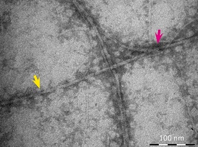
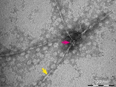
Figure 2.Fibril formation of HFIP treatment of Aβ40 (left) and Aβ42 (right). The magenta arrow points to the nucleation centers where aggregation starts. The yellow arrow indicates the fibrils which are formed as long thin helical structures with regular twists.
Early detection of AD
AD researchers are striving for better methods to detect onset of AD. To date, it has only been possible to detect AD during late stages, when significant damage to the brain has already occurred. Early detection of beta-amyloid in cerebrospinal fluid can be achieved through a lumbar-region puncture as well as by performing a positron emission tomography (PET) scan. However, both of these detection methods are expensive and not readily available in all global locations.
Efforts are ongoing for early identification of beta-amyloid build-up in the eyes of patients as a possible biomarker [5]. The peptide build-up begins 15 to 20 years before the onset of Alzheimer's symptoms. A study presented at the Alzheimer’s Association International Conference 2014 shared results showing a correlation between the levels of Aβ build-up in eyes and Aβ build-up in the brain [6].
Researchers continue to explore and gain a better understanding of the onset and possible treatments for AD.
References:
[1] Cárdenas-Aguayo, M del C, et al., “Physiological role of amyloid beta in neural cells: The Cellular Trophic Activity,” Neurochemistry, Chapter 9, 953-978, 2014.
[2] Zhefeng Gu, et al., ”Alzheimer's Aβ42 and Aβ40 peptides form interlaced amyloid fibrils,” Journal of Neurochemistry, 126:305-311, 2013. [PMID: 23406382]
[3] Stine Jr., WB, et al., “In vitro characterization of conditions for amyloid-β peptide oligomerization and fibrillogenesis,” Jour Biological Chemistry, 278:11612-11622, 2003. [PMID: 12499373]
[4] Pachahara, SK, et al., “Hexafluoroisopropanol induces self-assembly of β-amyloid peptides into highly ordered nanostructures,” J Pept Sci, 18:233-241, 2012. [PMID: 22252985]
[5] Frost, S, et al., “Ocular biomarkers for early detection of Alzheimer's disease,” J Alzheimers Dis, 22:1-16, 2010. [PMID: 20847434]
[6] Yen Ying Lim, et al., “Evidence of neuroinflammation in the retina in presymtomatic alzheimer’s disease,” Alzheimer’s & Dementia, 10:429, 2014.
Image:SEM of alzheimer's disease culture cells, Bachem
Related Products from: Bachem