BioLegend, Inc
Senior Product Manager
In defending the human body from invading pathogens, the immune system has to walk a fine line between reacting to foreign antigens and over-reacting to foreign antigens. To help modulate this response the immune system employs a subset of T-cells known as regulatory T cells (Tregs) that specialize in suppressing immune responses. Tregs play a critical role in balancing the activation of the immune system and immunological tolerance. Within the last decade there has been a great deal of effort to better understand the role of Tregs in the immune response. However, despite all the advances, there are still significant hurdles to overcome when studying this cell population. Most of these challenges are due to the fact that Tregs represent a very small population in any given tissue. But the development of efficient and innovative reagents for cell identification and isolation, along with the use of more standardized methodologies, are improving experimental results and the understanding of how Tregs function.
Detecting Tregs: intracellular vs surface markers
The history of Tregs goes back more than four decades. From 1969 to 1971, two groups described the suppressor capacity of thymus-derived cells [1]. However, the field advanced slowly due to the lack of a defined phenotype. Interest resurfaced more than 25 years later, with the description of CD25 and Foxp3 as suitable markers to identify these suppressor T cells, then renamed Tregs. The most specific Treg marker identified thus far is Foxp3, which is an intracellular transcription factor.
Here are some tips for Treg detection both intracellularly and via surface markers:
1) Flow cytometry can be used to isolate Tregs, however, this technique will only work if using surface markers to isolate the cells (unless the downstream application is compatible with fixed, dead cells).
2) In addition to Foxp3 there are other surface markers that can be used to avoid intracellular staining. For example, low or negative expression of CD127 (IL-7Ralpha) correlates with high expression of Foxp3 in human Tregs [2]. In mice, FR4 (Folate Receptor 4) can be used as well as CD4 and CD25 as detectable surface markers [3]
3) Human OMIPs (Optimized Multicolor Immunofluorescence Panel) have been reported to be used with flow cytometry to detect Tregs [4].
4) Intracellular staining requires fixation and permeabilization of the cells. In addition, the buffering system used will impact the quality of the results. Transcription Factor Buffer set from BioLegend was specifically developed for flow cytometry intracellular staining applications and provides superior signal-to-noise ratios when detecting Tregs.
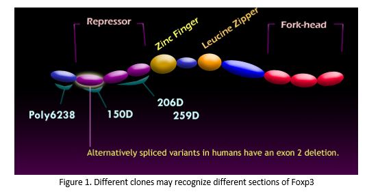
There are also several antibody clones available for Foxp3 (as well as many of the molecules mentioned above, see fig. 1) so depending on your particular experimental set up you may prefer one clone over another and it may take some experimentation to find the one that works best for you [5,6].
Nanobodies, the ultimate revolution in Treg recovery
Sample preparation is a critical step in the study of Treg cells; high quality data starts with a high quality sample. A frequent problem when working with Tregs, and many other cells, is that they die during the sample preparation. One mechanism that has been shown to affect Treg cells is the so called NAD-induced cell death (NICD).
The enzyme ARTC2 is highly expressed in Tregs and is also critical in the NICD process. By blocking this enzyme in vivo, the recovery of Tregs from mouse tissues increases dramatically [7]. BioLegend recently introduced a Nanobody that specifically blocks the enzyme ARTC2. A Nanobody is an antibody fragment consisting of a single monomeric variable domain engineered from a camelid heavy-chain only antibody, and has a molecular weight of only 15 kD (as compared to traditional antibodies such as IgG possess both heavy and light chains and typically have a molecular weight of 150 kD). Because of their size nanobodies can access sites that are not accessible to conventional antibodies (like catalytic sites of enzymes as shown in fig. 2).

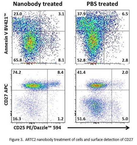
Damaged cells release NAD+ (Nicotinamide Adenine Dinucleotide). Free NAD+ binds cells expressing the enzyme ARTC2. ARTC2 then transfers ADP-ribose onto proteins like P2X7. This activates P2X7 for ion transport and causes downstream effects like cleavage of L-selectin (CD62L) and CD27, and flipping phosphatidylserine from the inner side of the membrane to the outer side. This initiation of apoptosis is termed “NAD+-induced cell death”, or NICD. Injection of mice with the nanobody blocks ARTC2 function, preventing apoptosis and shedding of surface markers, facilitating the recovery of live Tregs.
The following data illustrates that when mice receive a dose of an ARTC2 nanobody, the recovery of live cells increases dramatically. Their surface phenotype is also preserved, as shown by expression of CD27 (Fig. 3) and L-Selectin (not shown).
In the end
Optimizing your Treg research can be challenging as there are several factors to consider. But keeping these tips in mind as you plan your sample preparation and flow cytometry staining will help you maximize your results.
References
[1] Shevach E.M. The resurrection of T cell-mediated suppression. J Immunol. 186:3805-7, 2011. [PubMed ID: 21422250]
[2] Liu W. et al. CD127 expression inversely correlates with FoxP3 and suppressive function of human CD4+ Treg cells. J Exp Med. 203:1701-11, 2006. [PubMed ID: 16818678]
[3] Yamaguchi T. et al. Control of immune responses by antigen-specific regulatory T cells expressing the folate receptor. Immunity. 27:145-59, 2007 [PubMed ID: 17613255]
[4] Biancotto A. et al. OMIP-004: in-depth characterization of human T regulatory cells. Cytometry A. 81:15-6, 2012. [PubMed ID: 22052639]
[5] Law J.P. et al. The importance of Foxp3 antibody and fixation/permeabilization buffer combinations in identifying CD4+CD25+Foxp3+ regulatory T cells. Cytometry A. 75:1040-50, 2009. [PubMed ID: 19845018]
[6] Grant J. et al. Validated protocol for FoxP3 reveals increased expression in type 1 diabetes patients. Cytometry B Clin Cytom. 76:69-78, 2009. [PubMed ID:18690669]
[7] Koch-Nolte F. et al. Single domain antibodies from llama effectively and specifically block T cell ecto-ADP-ribosyltransferase ART2.2 in vivo. FASEB J. 21:3490-8, 2007. [PubMed ID: 17575259]
Image: BioLegend
Related Products from: BioLegend