How PCR Works
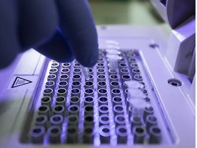 PCR uses repeated cycles of denaturation, annealing, and extension to exponentially create copies of target DNA sequences.2 In the denaturation phase, the DNA is heated to a temperature high enough to disrupt the hydrogen bonds linking complementary base pairs, opening the double helix into two distinct single strands. 95°C is the most common benchmark for this stage, but protocols can range from 92-98°C.2,4 The reaction then proceeds to the annealing phase, where the temperature is rapidly reduced to a range, usually between 55°C and 72°C, that permits primers to anneal to the exposed DNA templates strands.<sup>2</sup> This broader range is because optimal annealing temperature depends on the physical and chemical properties of the primers being used, which in turn varies depending on the template sequence being targeted.
Finally, the temperature ramps up to the 68°C to 80°C range for the extension phase, although scientists most commonly use 72°C. This discourages further primer annealing and promotes DNA polymerase activity, leading to the synthesis of two new strands complementary to the existing templates.2 This temperature cycling (thermocycling) then repeats, with the number of strands doubling each time, until the DNA product yield is sufficient for detection. Most PCR protocols entail 30-35 cycles, but 40 cycles is not uncommon for low concentration templates.2,4 Going beyond 40 cycles tends to reduce amplification efficiency due to reagent depletion, reduced enzyme activity, and excessive self-annealing.2
The Core Ingredients to a PCR Reaction
Fundamentally, PCR requires four key ingredients: the DNA template, primer sequences, additional deoxyribonucleoside triphosphate (dNTP) molecules, and a DNA polymerase. The template provides something to amplify, primers enable targeted amplification, the polymerase facilitates DNA replication, and the dNTPs provide the building blocks for this process.4 Before thermocycling, these ingredients are placed into a buffered water solution containing the ions, monovalent salts, and divalent cations required for polymerase activity.4
The precise composition of the buffer can vary and largely depends on the properties of the selected DNA polymerase. Accordingly, commercially available polymerases are almost always accompanied by a buffer solution tailored for its optimized performance. Common buffer ingredients include Tris (Tris-HCl but sometimes Tris-SO4) to control pH, K+ (usually as KCl) to promote primer annealing, and Mg2+ (typically as MgCl2) to facilitate polymerase activity.4While commercial products often provide pre-made buffer solutions with these aforementioned components, the magnesium solution will often be provided separately, letting researchers control final Mg2+ concentrations within the reaction. This is important as while too little magnesium leads to inactive polymerases, too much may increase non-specific amplification.4,5
How PCR Has Developed
While PCR has proven to be a transformative technology, it was not without its limitations. In particular, the ability to analyze the end-product only after amplification makes PCR unsuitable for quantitation. In response to this limitation, scientists developed quantitative PCR (qPCR), also known as real-time PCR, in the early 1990s.6 qPCR operates using the same denaturation, annealing, and extension cycles as PCR. However, it introduces a label that produces fluorescent signal relative to product amplification. Reading this fluorescence after every cycle lets researchers ascertain the amount of amplification required to obtain a particular amount of DNA and thus quantify target sequences relative to each other or to a standard curve.1,8 Since its invention, qPCR has become the gold standard for quantifying nucleic acids, and has gained widespread adoption, particularly for studying mRNA.1,7
The semi-quantitative nature of qPCR still offered room for improvement, leading to the development of digital PCR (dPCR). Here, the sample is partitioned using microwells into individual molecule-level portions, with amplification performed on all microwells simultaneously.8,9 Each well will return a “yes” or “no” signal depending on whether the target sequence was present, and then the proportion of “yes” wells is used to algorithmically calculate DNA concentration. dPCR does not offer a real-time window into DNA amplification, but provides absolute quantification capabilities.8,9
From relatively humble beginnings, PCR and all its subsequent iterations have quickly become a staple of the life sciences, biomedical research, clinical diagnostics, and more. Yet the PCR story is far from finished. So long as scientists continue to require sequencing information, PCR technology will continue to be in demand.
1. Zhu H. et al., “PCR past, present and future.” Biotechniques. 2020;69(4):317-325.
2. Khehra N. et al., “Polymerase Chain Reaction (PCR) [Updated 2025 Jul 7].” In: StatPearls [Internet]. Treasure Island (FL): StatPearls Publishing; 2025 Jan-.
3. Valones M.A. et al., “Principles and applications of polymerase chain reaction in medical diagnostic fields: a review.” Braz J Microbiol. 2009;40(1):1-11.
4. Wages J.M. Jr. “POLYMERASE CHAIN REACTION.” Encyclopedia of Analytical Science. 2005:243–50.
5. Lorenz T.C. “Polymerase chain reaction: basic protocol plus troubleshooting and optimization strategies.” J Vis Exp. 2012;(63):e3998.
6. Kubista M. et al., “The real-time polymerase chain reaction.” Mol Aspects Med. 2006;27(2-3):95-125.
7. Raso A., Biassoni R. “Twenty years of qPCR: a mature technology?” Methods Mol Biol. 2014;1160:1-3.
8. Quan P.L. et al., “dPCR: A technology review.” Sensors (Basel). 2018;18(4):1271.
9. Morley A.A. “Digital PCR: A brief history.” Biomol Detect Quantif. 2014;1(1):1-2.