Salk researchers have developed a new imaging method that lets them monitor actin, which are fine skeleton-like filaments within cells. With the new method, the team has been able to home in on how actin helps mitochondria divide in two. The work, published today in Nature Methods, could provide a better understanding of mitochondrial dysfunction.
"Actin is the most abundant protein in the cell, so when you image it, it's all over the cell," says Uri Manor, corresponding author of the paper. "Until now, it's been really hard to tell where individual actin molecules of interest are, because it's difficult to separate the relevant signal from all the background."
Mitochondrial fission is the process by which these energy-generating structures, or organelles, divide and multiply as part of normal cellular maintenance; the organelles divide not only when a cell itself is dividing, but also when cells are under high amounts of stress or mitochondria are damaged. However, the exact way in which one mitochondrion pinches off into two mitochondria has been poorly understood, particularly how the initial constriction happens. Studies have found that removing actin from a cell entirely, among many other effects, leads to less mitochondrial fission, suggesting a role for actin in the process. But destroying all the actin causes so many cellular defects that it's hard to study the protein's exact role in any one process, the researchers say.
Search Antibodies Search Now Use our Antibody Search Tool to find the right antibody for your research. Filter
by Type, Application, Reactivity, Host, Clonality, Conjugate/Tag, and Isotype.
So, Manor and his colleagues developed a new way to image actin. Rather than tag all the actin in the cell with fluorescence, they created an actin probe targeted to the outer membrane of mitochondria. Only when actin is within 10 nanometers of the mitochondria does it attach to the sensor, causing the fluorescence signal to increase.
Rather than see actin scattered haphazardly around all mitochondrial membranes, as they might if there were no discrete interactions between actin and the organelles, Manor's team saw bright hotspots of actin. And when they looked closely, the hotspots were located at the same locations where the endoplasmic reticulum crosses the mitochondria, previously found to be fission sites. Indeed, as the team watched actin hotspots light up and disappear over time, they discovered that 97 percent of mitochondrial fission sites had actin fluorescing around them.
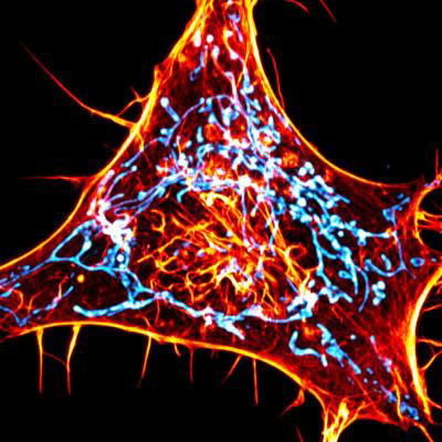
By altering the actin probe so that it attached to the endoplasmic reticulum membrane rather than the mitochondria, the researchers were able to piece together the order in which different components join the mitochondrial fission process. The team's results suggest that the actin attaches to the mitochondria before it reaches the endoplasmic reticulum. This lends important insight towards how the endoplasmic reticulum and mitochondria work together to coordinate mitochondrial fission.
Image: A cancer cell labeled for actin (red) and mitochondria (cyan). The scientists designed novel probes that specifically monitor interactions between actin and mitochondria. Image courtesy of Salk Institute/Waitt Advanced Biophotonics Center.