 According to a new study, scientists have used Positron Emission Tomography (PET) imaging to study brain inflammation following Zika virus infection in mice. The work was published last week online in Molecular Imaging and Biology from researchers at the US Army Medical Research Institute of Infectious Diseases (USAMRIID).
"Traditional methods of infectious disease research using animal models have provided limited information about disease progression until the study's endpoint, when investigators could analyze tissues from those animals. Imaging studies allow us to gather enhanced information through longitudinal studies of the same animal during the course of the infection," says senior author Thomas M. Bocan, Ph.D., of the USAMRIID.
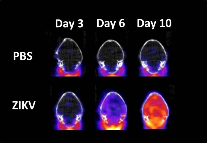
The researchers evaluated the ability and sensitivity of PET imaging, using a probe called [18F]DPA-714 to detect and quantify neuroinflammation in Zika virus-infected mice. Doing this they found that levels of Zika virus in the mouse brain increase from day 3 to day 10 post-infection. They also showed an increase in global brain neuroinflammation using [18F]DPA-714 PET imaging.
The work shows the role of global neuroinflammation in the progression of Zika virus affection and according to researchers, the first time that [18F]DPA-714 PET imaging is able to detect and quantify Zika virus-related neuroinflammation disseminated throughout the brains of infected mice.
"The future is bright for the application of imaging in infectious diseases," said Bocan. "Measures of virus and bacteria distribution and the consequences of infection can be assessed in real time in the same subject. In addition, treatment with countermeasures can be evaluated with a better understanding of the state of disease progression."
Image: Bottom row: The image shows representative PET/CT mouse brain images at days 3, 6 and 10 post-infection with Zika virus. Red area indicates neuroinflammation. Top row: Image represents PBS control mouse brain images. Image courtesy of US Army Medical Research Institute of Infectious Diseases.