A team of researchers have described the way in which a bacterial protein and a mineral combine to allow the bacterium to breathe when oxygen is not available. This information could lead to innovations in bio-based devices, such as sensors that diagnose disease or detect contaminants using a protein-material link as the basis of their design. The research was done at the Department of Energy's Lawrence Berkeley National Laboratory (Berkeley Lab) and published online in the Journal of the American Chemical Society.
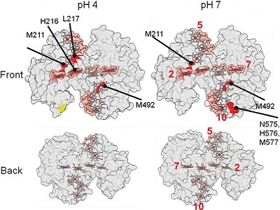
The protein used for this study is from a metal-reducing bacterium, Shewanella oneidensis, which can eat sugar and essentially breathe minerals in the absence of oxygen. While x-ray crystallography had previously been used to identify the structure of the protein, it wasn’t until the researchers used an X-ray-based technique known as “footprinting” that they were able to describe how the chemical connection between the bacterial protein and the nanoparticles composed of iron and oxygen is formed.
X-ray mass spectrometry footprinting allows scientists to precisely probe proteins and their surroundings and is currently only available at the ALS and Brookhaven's National Synchrotron Light Source II (NSLS-II).
In this technique, X-rays produce highly reactive hydroxyl radicals as they pass through a liquid solution that surrounds the protein. These radicals modify the protein, but the location where binding has occurred is protected from this change. Through mass spectrometry, researchers can analyze these chemical snapshots to determine how the protein is connected to the mineral.
One of the most surprising findings was that the protein binds rather weakly in this interaction. The researchers noted that this made sense given the function of the interaction-since the protein needs to transfer electrons to the mineral, the two do not need to be in contact for long.
The team plans to continue studying how this and similar proteins interact with various minerals and to find ways to use these interactions to make better electronic connections for more sensitive bioelectric sensors.
Image: Results from X-ray footprinting mass spectrometry (XFMS) experiments at Berkeley Lab's Advanced Light Source helped researchers identify where the protein binds with a mineral. The red areas indicate possible binding areas. Image courtesy of Berkeley Lab.