Clinical pathology laboratories are under increasing pressure to deliver accurate, reproducible results amid rising caseloads, workforce constraints, and growing expectations for faster turnaround times. These pressures are compounded by practical variables that begin long before an image is reviewed: delayed fixation, inconsistent tissue processing, sectioning differences, staining variation, scanner calibration, focus quality, and color reproducibility. For laboratories seeking to implement computational pathology safely and effectively, pre-analytic variability, staining and scanning inconsistency, workload pressure, and questions about AI reliability are not peripheral issues; they are central operational challenges that directly affect confidence in AI-assisted results and companion diagnostic readiness.
Reducing complexity and time
For today’s laboratories, the promise of digital and computational pathology is not simply technological advancement, but practical workflow support that helps pathologists and laboratory teams manage volume, complexity, and quality expectations. Modern platforms can integrate high-throughput image capture across brightfield, fluorescence, and multispectral modalities, with pattern recognition capabilities that help identify relevant tissue structures and cellular compartments.1 When implemented with appropriate laboratory controls and pathologist oversight, these tools can support more consistent quantification, such as immunohistochemistry (IHC) staining intensity analysis, while helping teams manage growing diagnostic and research demands.
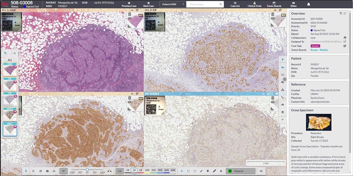
Digital pathology enables high-resolution imaging of slides, allowing users to zoom in and align images side-by-side for more effective comparison across tissue sections and IHC markers.
The urgency is clear: therapeutic development takes up to 15 years, requiring extensive assay development, analytical validation, and clinical validation—processes that are often conducted in silos and with limited integration.2 Diagnostic teams are frequently tasked with working backward from fixed clinical endpoints, often with limited visibility into the biological insights that informed the original therapeutic hypothesis.
Companion diagnostics to the rescue
Integrating diagnostics earlier in the development process, while fostering deeper collaboration across industry, academia, and healthcare, presents a significant opportunity to reduce costs, shorten timelines, and improve success rates.2 Reimagining drug development through companion diagnostics (CDx) is not merely a story of efficiency; it is essential to advancing human health and continuously accelerating idea-to-impact to enable access to lifesaving therapies.3
Despite their critical role in precision medicine, CDx development often trails behind therapeutic development. Too frequently, diagnostic strategies are introduced late in the process, sometimes only after a therapy has progressed into advanced clinical trials or even nearing regulatory submission. This misalignment creates downstream bottlenecks, forcing sponsors to reconcile diagnostic and therapeutic data under compressed timelines.
The consequences are significant. Late-stage integration can delay regulatory approvals, increase development costs, and introduce unnecessary risk, particularly when additional validation or bridging studies are required to align the diagnostic with the therapeutic. It also limits the ability to fully optimize patient selection strategies during earlier trial phases, potentially affecting clinical outcomes and overall program success.
Clinical pathology laboratories are essential to the success of companion diagnostics because they control many of the conditions that determine whether an assay can perform reliably in real-world use. Laboratories manage specimen quality, define and follow tissue-handling protocols, support analytical and clinical assay validation, monitor reproducibility, and translate validated approaches into routine practice. Their role is especially important as CDx programs move from controlled development settings into diverse clinical environments, where differences in specimen type, fixation time, staining platforms, scanner configuration, and workflow maturity can affect performance. By ensuring that tissue quality, assay execution, image generation, and interpretation are consistently governed, pathology laboratories help determine whether CDx approaches can scale from promising development programs to dependable clinical implementation.
Harnessing the benefits of artificial intelligence
AI-enabled companion diagnostics have the potential to support diagnostic accuracy, improve consistency, and reduce workload by assisting with tasks such as quantification, pattern recognition, triage, and workflow optimization.4 However, AI should be understood as a clinical decision-support tool rather than a replacement for pathologist expertise. Pathologists remain central to determining whether an AI output is clinically meaningful, whether it aligns with morphology and case context, and how it should inform patient-impact decisions. In this model, AI helps extend the reach and consistency of expert practice, while clinical judgment continues to rest with the pathologist.
This pathologist-led role extends beyond interpretation. Successful AI implementation depends on pathology-led validation, governance, quality oversight, and continuous performance monitoring. As laboratories process growing volumes of slides, AI models must be evaluated against the real conditions in which they will be used, including variation in fixation, staining, slide preparation, scanner performance, image quality, and patient and specimen diversity. Without this oversight, AI outputs may appear precise while still reflecting technical artifacts rather than clinically relevant biology.
Standardization of tissue processing
The reliability of computational pathology begins well before a slide is digitized. Pre-analytic variables such as delayed fixation, prolonged or inconsistent cold ischemia time, under- or over-fixation, tissue processing conditions, section thickness, microtomy artifacts, tissue folds, incomplete drying, and staining variation can all influence tissue morphology and biomarker expression. For example, delayed fixation may affect antigen preservation, inconsistent section thickness may alter apparent staining intensity, and staining variation can shift the visual features used for algorithmic classification or quantification. While experienced pathologists can often recognize and account for some of these variations during visual assessment, AI algorithms may be more sensitive to subtle differences that alter image characteristics. As computational pathology becomes increasingly integrated into research and clinical practice, standardized tissue processing protocols are essential to ensure consistency, reproducibility, and confidence in downstream analyses.
Standardization also plays a critical role in enabling multi-site studies, companion diagnostic development, and global clinical trial programs. When tissue preparation practices vary significantly across institutions, distinguishing true biological signals from technical artifacts becomes increasingly challenging. Establishing harmonized protocols, documented acceptance criteria, and quality metrics helps create more consistent datasets, enabling AI models and CDx assays to be trained, validated, and deployed across diverse healthcare environments with greater reliability.
Slide scanning quality control
High-quality whole slide images are fundamental to successful digital pathology workflows.5 Variability introduced during slide scanning—including scanner calibration differences, focus issues, illumination variation, color differences, resolution settings, image compression, and artifacts such as blur, striping, or out-of-focus regions—can influence both pathologist interpretation and algorithm performance. Even subtle inconsistencies that may not be immediately apparent during routine review can affect quantitative image analysis and AI-driven assessments, particularly when models are deployed across scanners, sites, and staining environments that differ from those used during development.
Implementing robust slide scanning quality control measures helps ensure that digital images accurately represent the underlying tissue. Routine scanner calibration, objective image quality assessments, color management, focus checks, and standardized operating procedures can reduce technical variability and support more reproducible results. As laboratories scale digital pathology operations, maintaining consistent image quality across scanners, sites, and time points becomes increasingly important for both clinical decision-making and AI readiness.
Impact of pre-analytic variability on AI models
AI models learn from patterns present within their training data and are therefore inherently influenced by the quality and consistency of the images they analyze. Variability introduced during tissue collection, processing, staining, or scanning can alter image features in ways that affect model performance, particularly when algorithms are deployed in environments that differ from those represented in development datasets. For example, delayed fixation may change antigen preservation, inconsistent section thickness may alter staining intensity, scanner focus issues may obscure cellular detail, and color differences across sites may shift the image features that an algorithm uses for classification or quantification. These differences can be especially consequential for CDx applications, where assay reproducibility and patient selection depend on consistent performance across real-world laboratories.
Addressing pre-analytic variability requires a comprehensive approach that combines laboratory standardization, diverse training datasets, rigorous validation across multiple institutions and populations, and pathologist-led review of algorithm performance. Models that are developed and tested using data generated under a range of real-world conditions are more likely to demonstrate robust performance when deployed at scale. Ultimately, reducing pre-analytic variability strengthens trust in AI outputs and helps ensure that clinical decisions are driven by meaningful biological insights rather than technical inconsistencies.
Harmonizing pathology workflows for AI readiness
Preparing pathology laboratories for AI adoption requires more than implementing digital scanners or deploying advanced software. AI readiness depends on the harmonization of workflows across the entire pathology continuum, from specimen acquisition and tissue processing through image generation, analysis, reporting, data management, and post-deployment monitoring. Each step contributes to the overall quality and consistency of the data that serves as the foundation for computational pathology applications, and each step requires clear accountability within the laboratory.
Organizations that proactively align processes, quality standards, technology infrastructure, and pathologist-led governance are better positioned to realize the full value of AI-enabled diagnostics. Harmonized workflows support reproducibility, facilitate regulatory compliance, and enable more efficient deployment of algorithms across laboratories and healthcare networks. By embedding standardization, quality management, and clinical oversight throughout the pathology workflow, institutions can create a scalable foundation for innovation while maximizing confidence in AI-assisted decision-making.
Benefits of end-to-end solutions
The advantages of an AI-powered digital pathology lab are further amplified when delivered through a unified, end-to-end solutions provider. By integrating hardware, software, workflows, and data management within a single cohesive ecosystem, laboratories can significantly reduce operational complexity, enhance interoperability, and minimize workflow disruptions.6
This integrated approach enables seamless data flow from image acquisition through analysis and reporting, accelerating turnaround times while improving consistency and data integrity. A standardized platform also simplifies validation processes, supports regulatory alignment, and reduces the burden on internal IT infrastructure, ultimately driving greater efficiency, scalability, and confidence in results.
As computational pathology continues to evolve from a promising innovation to a core component of precision medicine, success will depend not only on the sophistication of AI algorithms, but also on the quality, consistency, and clinical governance of the data that underpin them. Standardized pre-analytic processes, robust scanner and image quality controls, harmonized workflows, and pathologist-led validation create the foundation necessary for AI to deliver reliable, reproducible, and clinically meaningful insights.
For pathology leaders, the call to action is practical and immediate: standardize tissue handling and fixation protocols; define quality criteria for sectioning, staining, scanning, and image review; implement scanner and image quality control programs; validate AI models across diverse sites, specimen types, staining conditions, scanners, and patient populations; and involve pathologists early in AI and companion diagnostic governance. These priorities should be treated not as downstream implementation details, but as core requirements for trustworthy AI-enabled diagnostics. By connecting workflows from specimen to insight and aligning expertise across laboratory, clinical, technology, and biopharmaceutical stakeholders, pathology teams can help ensure that AI-enabled CDx solutions are trustworthy, reproducible, and ready for real-world clinical implementation.
References
1. Locke D, Hoyt CC. “Companion diagnostic requirements for spatial biology using multiplex immunofluorescence and multispectral imaging.” Frontiers in Molecular Biosciences. 2023;10:1051491. doi:10.3389/fmolb.2023.1051491.
2. Chatterjee B, Steiner R, Kaul G. Industry Perspective – What does Industry Need to Accelerate Drug Product and Process Development? Pharm Res. 2024 Jan;41(1):7-11. doi: 10.1007/s11095-023-03604-y. Epub 2023 Oct 11. PMID: 37821765; PMCID: PMC10810959.
3. Wu Y, Xue R, Luo X, Liao J, Zhang Z, Deng J, Liu T, Li X, Chen ZS, Yin M. Companion Diagnostics in Clinical Therapy: Current Applications and Future Directions. MedComm (2020). 2026 Mar 1;7(3):e70638. doi: 10.1002/mco2.70638. PMID: 41777248; PMCID: PMC12950518.
4. Jeong J, Kim S, Pan L, Hwang D, Kim D, Choi J, Kwon Y, Yi P, Jeong J, Yoo SJ. Reducing the workload of medical diagnosis through artificial intelligence: A narrative review. Medicine (Baltimore). 2025 Feb 7;104(6):e41470. doi: 10.1097/MD.0000000000041470. PMID: 39928829; PMCID: PMC11813001.
5. Leica Biosystems. “How Whole Slide Imaging Is Changing Pathology.” https://www.leicabiosystems.com/us/life-sciences-and-research-solutions/application/how-whole-slide-imaging-is-changing-pathology/
6. Fraggetta F, L’Imperio V, Ameisen D, et al. Best Practice Recommendations for the Implementation of a Digital Pathology Workflow in the Anatomic Pathology Laboratory by the European Society of Digital and Integrative Pathology (ESDIP). Diagnostics. 2021;11(11):2167. doi:10.3390/diagnostics11112167.