Flow cytometry is a technique that’s continually evolving. Improvements to instrumentation, reagents, and software have all been key in enabling researchers to generate increasingly detailed profiles of individual cells. However, while the value of performing multiparametric analysis at a single-cell level is widely recognized, there are still those who regard flow cytometry as a complex technique that is best left to the experts. Here, we explain why this is no longer the case and discuss recent developments that have led to flow cytometry being used for a growing number of applications.
Almost any researcher can perform basic flow cytometry
According to Bob Smith-McCollum, director of global marketing for flow cytometry at Luminex, flow cytometry was once considered a ‘dark art’ practiced by experts who were part life scientist and part engineer. Despite being avidly adopted by immunologists keen to reap the benefits of single-cell analysis, it was only those with a reasonable level of expertise who were able to generate meaningful data. Moreover, to achieve results, they were confined to using large, bulky systems housed in shared equipment core labs. “Fortunately, times have changed,” notes Ken Lau, Ph.D., senior technical marketing manager at BioLegend, “and all components of flow cytometry have evolved such that researchers with limited knowledge of the technology can now comfortably use relatively complex multicolor panels in their research.” Mike Blundell, Ph.D., field marketing specialist at Bio-Rad, agrees, noting that advances in reagents, instrumentation, and software have all helped encourage more researchers to begin running flow cytometry experiments.
Smaller, more powerful instrumentation
A striking difference between early flow cytometry and modern forms of the technique is the size of the instrumentation. Lauren Jachimowicz, application development scientist at Agilent, points out that one of the main reasons flow cytometry has been able to reach into new and developing fields is that benchtop flow cytometers can be positioned in a small lab space rather than requiring a dedicated facility. “Our new NovoCyte Penteon has a footprint of just 62 cm x 46 cm,” she says, “meaning it can easily be placed in a cell culture lab. Not only does this help to streamline flow cytometry workflows but it also eliminates the need to transport precious sample material that may oftentimes be irreplaceable.”
Another important differential is the increased number of lasers, filters, and detectors that have contributed to larger panel sizes. “The types of detectors and fluidics have also evolved to meet demand for larger panels,” explains Blundell, “along with the level of control that can be applied to the various parts of a flow cytometer, and the speed at which samples can be acquired.” Modern instrumentation also encompasses spectral cytometers that allow for detection of more readouts from a single laser (for example, the Cytek® Aurora has up to five lasers, three scatter channels, and 64 fluorescence channels) and imaging flow cytometers that add quantitative microscopy imaging to every cell (for example, the Amnis® FlowSight® and ImageStream® from Luminex).
Dyes with unique spectral signatures
As is the case for almost any technology, instrumentation and reagents have evolved hand in hand. Lau observes that having more fluors with unique properties to choose from helps researchers create more balanced panels or even expand the number of parameters to be analyzed, while Blundell highlights that fluorophore characteristics have improved to incorporate narrower excitation and emission spectra, longer Stokes shifts, greater stability and better lot-to-lot reproducibility. It would, of course, be impossible to cover all the available fluors here, but newer options are BioLegend’s suite of fluorophores that work ideally with spectral cytometers (including APC/Fire™ 810, one of the first flow cytometry options to extend into the far red with its emission spectra, and fluors such as Spark Blue™ 550 and Spark NIR™ 685 that fill gaps between existing fluors), and Bio-Rad’s StarBright dyes, a family of extremely bright fluorescent nanoparticles developed specifically for flow cytometry that allow detection of rare cells and low antigen density populations.
Intuitive software for acquisition, analysis, and reporting
A further contributing factor to the rising uptake of flow cytometry is the development of more user-friendly software for data acquisition, analysis, and reporting. Researchers can now run flow cytometry experiments without the need for specialized training and can leverage features like automatic compensation, auto-gating, and a broad range of display options for easier data capture and downstream processing. “Since multiparameter analysis has become increasingly complex, manual gating has become more time-consuming and difficult to perform,” notes Jachimowicz. “However, with advances in machine learning, automated analysis is helping to eliminate errors in manual gating and provide more reliable identification of complex patterns across markers and cells.”
“Artificial intelligence has applicability across the entire flow cytometry process,” adds Blundell. “At one end, it can be used to improve fluor selection and reduce trial and error during panel building, while at the other it can be employed to improve data analysis. Gating and analyzing high parameter panels can be a laborious and somewhat subjective process, with different cytometrists preferring different gating strategies. Additionally, high parameter data does not benefit from two color dot plots and sequential gating and current methods like SPADE analysis or tSNE plots are not always the most informative. The advancement of machine learning-driven auto-gating based on large historical datasets will remove any potential bias or subjectivity in data analysis to increase throughput, improve data quality, and free up time for researchers.”
Supporting new applications
Common applications of flow cytometry include immunophenotyping, cell cycle analysis, the study of apoptosis, and biomarker discovery and diagnostics, yet the technology is branching into many other research areas as well. For instance, flow cytometry is now used to analyze other regulated cell death pathways such as autophagy and necrosis, and it has recently found itself at the center of research for COVID-19 where it has been used to determine immune responses, recovery, and antibody levels through cellular and bead assays. “The multiparameter data acquired, at speed, has certainly helped with research into the basis of the disease and will be an important tool for determining vaccine efficiency,” says Blundell.
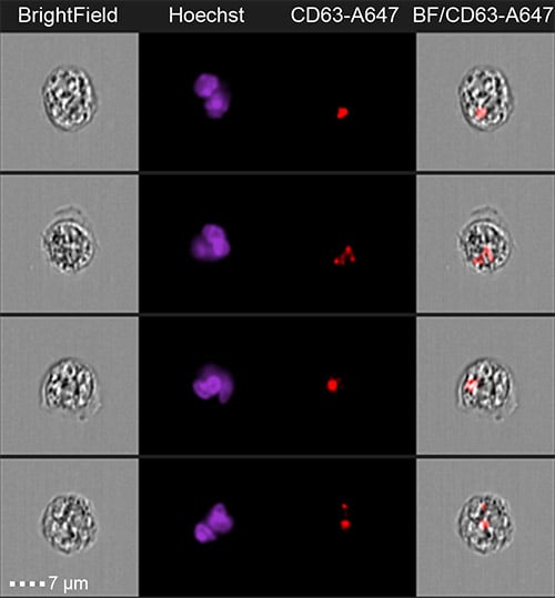
Image: Jurkat-derived extracellular vesicles labeled with anti-CD63 Alexa Fluor 647 internalized by neutrophils from human blood. Images collected at 60x magnification on an Amnis ImageStream®X Mk II imaging flow cytometer from Luminex.
A further novel use of flow cytometry has been to study extracellular vesicles—small particles that are released under normal physiological conditions as well as during various disease states and that have been shown to transfer biological molecules, potentially transmitting signals. “Analyzing small particles such as exosomes has been made possible by a combination of improved instrument sensitivity, better antibody reagents, and the development of exceptionally bright dyes,” says Lau. “These types of studies can provide valuable information to support early detection of disease and can also enable mechanistic insights into cancer and autoimmune conditions.”