Using the wrong antibody reagents for flow cytometry can be disastrous. A poor choice can result in wasted time, money, and precious sample material, and may lead to the publication of incorrect or irreproducible data. Although the risk of these outcomes can be reduced, doing so does not simply involve selecting antibodies that are purported to be suitable for flow. Researchers should consider factors such as the antibody format, the vendor’s validation method, and the nature of the antigenic target to increase the likelihood of flow cytometry success.
“There are many common mistakes when choosing antibodies for flow cytometry,” reports Mike Blundell, product manager, flow cytometry at Bio-Rad. “These include picking an antibody that recognizes the wrong epitope, such as a product targeting an intracellular epitope when the intention is to avoid permeabilizing the cells; or selecting the wrong clone, for instance assuming an antibody that has been used in another application will work for flow. It is also possible to choose an inappropriate marker; Bio-Rad’s online cell marker selection tool can help researchers to avoid false positives that occur as a result of detecting multiple cell types.”
“Another factor that researchers should consider when selecting an antibody for flow cytometry is whether to use a directly conjugated or unconjugated product,” adds Wayne Austin, Ph.D., senior scientist, flow cytometry at Abcam. “Directly conjugated primary antibodies are by far the preferred format for flow, allowing simultaneous use of multiple antibodies raised in the same species and affording increased experimental complexity. Furthermore, having direct conjugates simplifies the assay protocol, reducing the potential for mistakes to occur during sample preparation. If the desired antibody-fluorophore combination is unavailable, Abcam offers a range of conjugation-ready antibody formulations that have been validated for flow, as well as a portfolio of easy-to-use conjugation kits.”
Researchers should also choose a monoclonal antibody, ideally a recombinant one, for their flow cytometry experiment whenever possible, notes Austin. This is because polyclonals are more likely to cross-react with proteins other than the target and frequently exhibit variable batch-to-batch performance. “Monoclonals, especially recombinant ones, are consistent and reproducible,” he says. “At Abcam, we’ve engineered recombinant versions of over 19,000 of our RabMAb® rabbit monoclonal antibodies, allowing researchers to benefit from enhanced specificity and sensitivity. Many of these products have been knockout validated using CRISPR/Cas9-modified cell lines and tested for use in flow.”
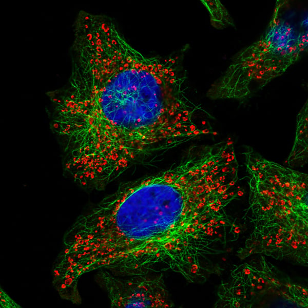
Image: HeLa cells stained with a recombinant rabbit monoclonal antibody to ATP5A conjugated to Alexa Fluor® 594 (red, ab216385), as well as a mouse monoclonal antibody to alpha Tubulin conjugated to Alexa Fluor 488 (green, ab195887). Nuclear DNA was labeled with DAPI (blue), and the image taken with a confocal microscope (Leica-Microsystems, TCS SP8). Image courtesy of Abcam.
According to Dr. Michael Fiebig, business development director, products and innovations at Absolute Antibody, there are a number of advantages to choosing recombinant antibodies for research, especially when it comes to flow cytometry. “In general, using recombinantly produced antibodies ensures true clonality, reducing the chance of off-target binding and delivering the highest possible batch-to-batch reproducibility,” he explains. “With regards to flow cytometry, recombinant technology allows the selection of new, engineered versions of an antibody that better match the experimental design and can improve readouts.

Image: Fc Silent represents a genetically engineered Fc domain containing key point mutations that abrogate binding of Fc receptors, reducing background staining in vitro and abolishing antibody directed cytotoxicity (ADCC) effector function in vivo. Image courtesy of Absolute Antibody.
To explain this concept, Fiebig describes the use of recombinant technology to prevent IgG antibody binding to Fcγ receptors, a well-known cause of high background in flow. “Many researchers opt to use antibodies that bind to FcγR, leading to additional staining of all cells that express these receptors,” he says. “For instance, isotypes such as mouse IgG2a/b and rat IgG2b can greatly increase background staining by binding macrophages, monocytes, or B-cells. Although you can address this issue by adding Fc-blockers or serum to your sample preparation, this can lead to incompatibilities with other reagents. As an example, following treatment with the most widely used mouse CD16/32 blockers you cannot stain for rat antibodies any more as the blocking agent is also a rat antibody. To address this problem we developed our Fc Silent™ antibodies. These are improved versions of classic clones that have been engineered to express a small set of mutations preventing FcγR binding. Our Fc Silent technology can be applied to any species and generates full-length antibodies that can replace conventional antibodies in flow cytometry staining protocols.”
With so many antibodies available, it can often be overwhelming to select a suitable product. This is where the level of detail provided by the vendor comes into play. “Checking the clone and the validated applications along with recommended antibody dilutions and staining protocols on the vendor website before using an antibody will help prevent poor or inappropriate staining,” says Blundell. “For example, Bio-Rad’s F4/80 antibody is one of the most well-characterized and extensively referenced mouse macrophage markers for staining spleen sections. However, for flow cytometry, spleen samples must be mechanically homogenized and treated with collagenase to release the red pulp macrophages; failure to do this can result in misleading data.”
Christopher Manning, group leader (flow cytometry) at Cell Signaling Technology, notes that for an antibody to be approved for flow, it should be properly validated by both the vendor and the end user prior to hypothesis-driven research. “I would recommend researchers first look for antibodies that have already been validated in flow, and then further validate the performance of those products in their own model,” he says. “Relevant positive and negative expression controls are especially important to flow cytometry since the researcher sees only the raw fluorescence detected by the instrument—there is no localization, molecular weight, or other indication that an antibody is performing as expected.”
At Cell Signaling Technology, controls for antibody validation include a positive cell line versus a knockout or negative cell line (for antibodies targeting total protein), or treatment conditions that promote or inhibit defined modifications (for antibodies targeting post-translational modifications). The company also provides detailed, application-specific protocols, yet Manning stresses the importance of validating any antibody in the exact protocol that will be used in-house. “Confirming specificity in your particular experiment and cell type is essential,” he says. “Moreover, should it become necessary to switch reagents or conditions, antibody staining should be revalidated—a simple one-day experiment to confirm specificity can save years of lost research when it becomes apparent that an antibody is not performing as assumed.”
While flow cytometry is benefiting from improved antibody reagents and more rigorous validation by both vendors and researchers, there is still room for improvement, particularly when it comes to streamlining the selection of appropriate antibodies for increasingly complex flow cytometry experiments. “I feel that the flow cytometry reagents market suffers from a race-to-the-bottom in terms of pricing, but with so many vendors competing over the same clones this is hardly surprising,” observes Fiebig. “The interest we are finding in our recombinant antibody products is testament to their usefulness in designing new flow cytometry experiments and improving well-established methods. The superior reproducibility, low background, and ability to streamline reagent panels that is offered by recombinantly produced reagents has begun to entice flow cytometrists toward a new way of thinking.”