High-content systems for screening, analysis, and imaging are powerful tools for life science research. These systems are typically designed for rapid data acquisition and image capture. In combination with new, more sophisticated assay types, their use in drug screening, toxicity testing, and disease modeling is rapidly expanding.
3D cell models have become a disruptive force in high-content screening technology. Instrument manufacturers are scrambling to upgrade their products to make the most of the enhanced structural and functional detail available in 3D cell cultures and organoids.
StemoniX is a life science company built on nano- and micro-manufacturing techniques from the semiconductor industry. It manufactures assay-ready, 2D and 3D plates for high-content screening based on the company’s engineering values of consistency and accuracy. “I’d say that one of the things that we’ve brought to the industry with our assay-ready plates is this idea of accurate cell structures,” explains Ping Yeh, StemoniX co-founder. According to Yeh, accurate cell structures modeled in three dimensions can produce a more biologically relevant model.
For example, Stemonix’ microHeart product uses cells formed into microfibers, similar to cells in their natural environment. That offers the ability to study the alignment of fibers and structures such as sarcomeres that are found in cells in the human body. “We’re encouraging cells in culture to interact with each other in unique and more physiological ways,” adds Yeh. In addition to their use in studying cellular structure, the platform may be used to identify markers of functionality as well as for drug toxicity investigations and visualization of electrochemical cellular processes.
One advantage of high-content screening with a product such as StemoniX’ assay-ready plates that more accurately model cellular biology is the ability to connect assay results with functionality. For example, according to Yeh, in gene expression analysis, high-content screening can provide confirming data for cell biology. “It opens a whole new world of possibilities for drug discovery. You can have a more empirical or experimental approach as opposed to one complicated by the behavior of animals.”
High-content screens using StemoniX’ platform can process dozens of plates in a single screen, all from the same lot of cells, offering consistency of results. One recent application of StemoniX technology is a study by the United States Army Medical Research Institute of Infectious Diseases evaluating the microBrain Assay Ready product in studies of infectivity with four viruses: Eastern equine encephalitis, Western equine encephalitis, Venezuelan equine encephalitis (VEEV), and Marburg virus (MARV). They also tested drug inhibition of viral infectivity against MARV and VEEV. They demonstrated that the virus strains could be easily titrated on the StemoniX platform, and that viral infection could be blocked using drugs.
Functional toxicology
Three-dimensional (3D) models have proven useful for interpreting complex biology of cells and tissues and have been used extensively in developmental biology and toxicity screening. Traditionally, cell cultures in 2D monolayers have been used for these studies. However, mechanical stress created by adhesion to an extracellular matrix or to other cells changes the processes of signal transduction and gene transcription. Three-dimensional cell cultures have more faithfully reproduced the functional activity of these cells than 2D cultures. Gene expression analysis has also shown that 3D cell culture triggers different patterns of gene expression.
Advances in high-content imaging have allowed the use of multiple fluorescent dyes to characterize cell responses and morphology.
Advances in high-content imaging have allowed the use of multiple fluorescent dyes to characterize cell responses and morphology. These advances include higher magnification and 3D analysis. Scientists at Molecular Devices have combined confocal high-content imaging with 3D image analysis to develop a high-throughput compound screen using liver spheroids made from iPSC-derived hepatocytes.
They used the ImageXpress® Micro Confocal High-Content Imaging System and analyzed the data using MetaXpress® companion software, which converts a stack of 2D images into 3D space. They found that 3D liver cell models accelerated in vitro toxicity assessment, delivering phenotypic readouts that allow screening for test compound effects on cell morphology and viability through a simple, continuous workflow. Different readouts were used to assess different aspects of cells such as spheroid morphology, integrity, viability, and toxicity effects.
In addition to improved 3D capabilities, ImageXpress Micro systems enable researchers to conduct various functional assays such as cardiac beating or calcium response analyses. Grischa Chandy, product manager for the high-content imaging line at Molecular Devices, explains that ImageXpress Micro systems allow capturing of a fast burst of data in an automated fashion, thus enabling visualization and measurements of hard-to-acquire responses. This allows users to perform high-content imaging as a functional assay for compound screening. “Most systems on the market can’t utilize the full speed of the camera, while our MetaXpress software combined with the ImageXpress Micro systems can take advantage of the camera’s very high frame rates, up to 100 fps. This allows both imaging and analysis of fast kinetic assays such as cardiac cell beating.”
Biocompare’s HCS Systems Search Tool
Find, compare and review HCS systems
from different suppliers Search
Proper imaging of a 3D cell model requires maintenance of its natural water-based environment, while focusing deep within the sample. Conventional microscopy using air objective lenses is subject to optical distortions for those deeper levels. A number of manufacturers have now introduced water immersion objectives to allow better visualization of the structures within a 3D sample.
Perkin-Elmer’s Opera Phoenix high-content imaging system combines a water immersion objective with its spinning disk confocal technology. A spinning disk confocal scanner works through rotating disks that have thousands of pinholes arranged in a spiral. Most of the incident light on the disk is reflected, but the holes create concentric arcs of excitation light on the sample. Fluorescent light from the specimen returns through the objective lens and pinholes and is relayed back to the detector. The entire field is scanned in the course of a single film exposure of the scientific complementary metal oxide semiconductor (sCMOS) camera, forming the confocal image at high speed.
Jacob Tesdorpf, director of high-content and detection instruments, discovery and analytical solutions for Perkin-Elmer, says that, “Microlenses allow a lot of excitation light to get deep in the tissue, and on the way back collect emission light, making two separate passes through the spinning disk.” Tesdorpf adds that the system can image up to four colors in parallel. Because those channels are separate, sensitivity is enhanced.
“Compared to older Opera instruments, we’ve seen great speed improvements depending on the type of assay you would do,” Tesdorpf notes. “You would certainly get a threefold, sometimes even bigger increase in speed just because of the advances in camera technologies.”
The trend toward more biologically relevant 3D high-content screening has driven advances in imaging technology, particularly in the area of microscopy. As the industry moves toward thicker 3D structures, there is more of a need to improve resolution in the Z dimension—or depth—of the image. GE Healthcare is anticipating that need with the latest product in its IN Cell series, the IN Cell Analyzer 6500HS. Compared to its previous model, the IN Cell Analyzer 6000, the HS (for High Sensitivity) is between two and five times more sensitive. The improvements include the latest sCMOS camera.
“You can do assays you couldn’t do before because you can see the dim structures in the sample,” says Will Marshall, product manager for high-content analysis with GE Healthcare Life Sciences Cell Analysis. As well, Marshall explains that the system can process more samples in the same amount of time. In addition to the sCMOS camera the 6500HS uses a line scan method for achieving confocal effect, rather than a spinning disk technology. “What this allows us to do is optimize the balance between speed and image quality in context of the specific experimental goal, which is not possible with other technologies such as a spinning disk,” Marshall adds. With its newer instruments GE is tackling several of the problems associated with extracting quantitative data from 3D cell culture samples.
In a recent study, GE and collaborators demonstrated the use of a tissue-clearing technique in combination with the GE IN Cell 6000 to more accurately assess drug effects in HepG2 spheroids. The clearing agent’s ability to mitigate light scattering allowed for better quantification of heterogeneous cell responses as a function of the spheroid local environment. Also key to the study’s success was the use of the IN Cell SmartScan feature, which reduced acquisition times by intelligently guiding the system to specific areas of interest in the sample.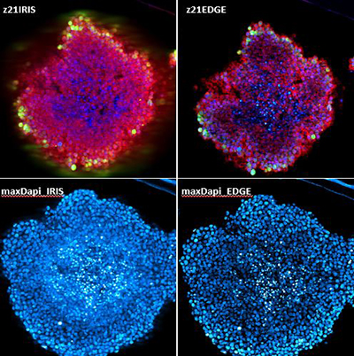
Image: All images are fluorescent microscopy images of the same sample. Sample is a tumor spheroid. Approximately 600 microns in diameter. maxDapi_EDGE is a maximum projection image of DAPI stained nuclei with EDGE confocal. maxDaPi_IRIS is a maximum projection image of DAPI stained nuclei with IRIS confocal (regular confocal). Notice that in comparison the EDGE image is much clearer. Z21EDGE is a single z plane from the same sample in 3 colors using EDGE confocal. Z21IRIS is the same plane in 3 colors using IRIS confocal. Again, notice that in comparison the EDGE image is much clearer. The “colors” or fluorescent labels are DAPI, calcein AM, and propidium iodide. Image courtesy of GE Healthcare Life Sciences.
Deeper analysis
Analysis software is another important tool for high-content screening, especially in three dimensions. Marshall emphasizes that pairing a high-content screening system with powerful image-analysis software is important to get the most out of a screen. GE’s IN Carta software is designed to work with high-quality images produced by its IN Cell analyzers. “We’ve made the analysis problem simpler by the way we structured this software, with the adoption of image-analysis algorithms and machine-learning techniques to solve problems.”
According to Othmar Pfannes, CEO of Genedata, traditional computer vision doesn’t scale well with the analytical complexity and sheer quantity of data produced by 3D model systems. They often require complex image-analysis procedure, with lengthy set up.
 Genedata’s Deep Learning technology uses convolutional neural networks (CNNs) to automatically analyze microscopy images in high throughput, simplifying image-analysis workflows. “The automated transfer of learned knowledge, input by a human expert, to a different experimental situation is now feasible. Producing quality results from a new experiment now takes just seconds, versus weeks typically required by manual optimization,” says Pfannes.
Genedata has applied its technology to a number of research projects. Those areas of application include quantification of phenotypic changes in a fibrosis assay using primary human fibroblasts, application of a deep-learning based similarity map to characterize differentiation of hMSCs into different lineages, and creating an efficient deep-learning workflow for routine high-content screening on many cell lines without requiring experts to tailor the image analysis for each line.
Image: Two cellular phenotypes (top) and the corresponding activation maps (bottom). Activation maps provide an understanding on which parts of the image are most influential for the classification by the CNN. Image courtesy of Genedata.
The availability of more biologically relevant model cell systems has radically increased the amount and speed of data that can be collected in high-content screening. Image-analysis technology is keeping pace with advances in cell biology, offering a novel look at function within deep structures resembling the native environment of cells. Those enhanced images, in turn, provide more variables for study of cell processes, effects of drugs, and toxicity.