Small animal imaging has a long history, originating with x-ray technology borrowed from the clinic. Other technologies, including three-dimensional x-ray (computed tomography, or CT), magnetic resonance (MR), as well as those based around radioactive tracers (such as positron emission tomography, or PET) and sound waves (ultrasound) have trickled down as well, further allowing researchers to probe the structure and molecular happenings of laboratory animals. Optical imaging, on the other hand, began its life in the preclinical world a couple of decades ago with the introduction of the IVIS (In Vivo Imaging System) by a predecessor of Revvity, and has been slowly trickling up to the clinic.
In this article, we look at the ways that light—the most widely used preclinical imaging modality—is being used to examine what goes on under the skin.
Optical modalities
Small animal optical imaging generally implies the detection of light emitted by molecular probes found in an intact animal. Probes range from conjugated antibodies or ligands to bacteria to genetically introduced proteins to labeled polymers, for example, and may be used to follow metastases, infection, small molecule or cellular biodistribution, or gastric clearance. Functional studies can be performed using probes that bind or are activated only under specific conditions.
Developed at a time when most often animals were sacrificed and evaluated by histopathology, the ability to follow optical signals over time “in the live animal was quite revolutionary,” remarks Alexandra De Lille, director of technical applications, in vivo imaging at Revvity.
In vivo optical imaging comes in two principle flavors: (bio)luminescence, which results from an enzymatic reaction, and fluorescence, in which light of one wavelength is captured by the probe and emitted at a different (usually longer, lower-energy) wavelength.
“We find the interest in both types of imaging now to be almost equal,” says Sean Gallagher, director, research and development at Analytik Jena US.
Other (specialized, hybrid) techniques—such as Cerenkov imaging, which looks at fluorescent signals generated from radionuclides, and photoacoustic tomography (PAT), which detects the sound emitted after a light pulse hits a probe—are sometimes considered to be forms of optical imaging, broadly defined.
Bioluminescence
Bioluminescence relies on the cleavage of a substrate (luciferin) by an enzyme (luciferase) in the presence of oxygen and ATP to produce light. In a typical experiment cells transfected with a luciferase construct are injected into a mouse as a xenograft under the skin. After the appropriate time and treatments, “all we have to do is inject luciferin intravenously or into the gut … It takes about five to ten minutes for the luciferin to circulate. And once the two interact then you get a photon that’s produced in that reaction as one of the byproducts,” explains Justin Jeffrey, manager of the UW Carbone Cancer Center Small Animal Imaging Facility at the University of Wisconsin in Madison.
Originally from the firefly, there are now systems derived from other insects, marine organisms, and bacteria, for example, as well as engineered versions, each with distinct characteristics such as kinetics, sensitivity, and wavelength.
Japanese scientists reported earlier this year, for example, a luciferase system created by directed evolution that is red-shifted and up to 1,000 times brighter than conventional systems. This enabled them to visualize and trace single cells inside moving mice, and to record bioluminescence from neurons deep inside a marmoset brain.
The conventional (green) luminescent signal is attenuated by tissue absorption. Yet “the reason why bioluminescence is so hot is because of its high signal to noise,” points out Andrew Van Praagh, lead applications scientist at Spectral Instruments Imaging. Unlike fluorescence, there is no external light introduced to the system, and no background to contend with. “The resulting sensitivity is equal or superior to most fluorescent probes.”
Fluorescence
Van Praagh points to three principle advantages of fluorescence: the ability to use multiple fluorophores at once in a mouse model (so long as they have distinct excitation and emission peaks); no steady state kinetics requirement (in contrast to luminescence)—you can take an image any time you want and will get the same intensity; and the ability to do ex vivo analysis— “it’s not enzymatically based, it doesn’t require oxygen, and it doesn’t require living cells like luminescence does.”
Penetration for fluorescent probes is in the range of several millimeters, “depending on how much you can get to your target,” says Joy Kovar, principal scientist at LI-COR Biosciences. That depth can be extended by using probes that excite and emit at longer wavelengths—into the far red and near-infrared (NIR) range—where autofluorescence, interference, and scattering from tissue is less.
Fluorescent protein probes “were first used to characterize cell epitopes and metabolic functions in vitro—in that situation there’s no tissue-associated attenuation, so they work just fine,” explains Van Praagh. 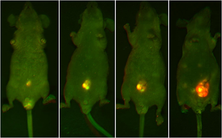 But GFP and RFP are of more limited use in a 2 cm-thick mouse, let alone a rat ten times the size. Fortunately, “there are a lot of body compartments that are superficial, in which case you can use RFP quite readily.” NIR dyes and conjugates are increasingly available, “and if you look in the literature there are NIR plasmid constructs that are going commercial.”
Image: Representative mouse highlighting the progressive increasing luminosity of an implanted lesion in the peritoneal cavity. Each image was captured using both GFP and RFP filters and then multiplexed. The intense fluorescent signal shown in the implanted tumor is significantly brighter than the surrounding tissue. Note the formation of metastatic lesions beginning to develop distal to the site of implantation and becoming visible within the abdomen in the final slide (solid white lines). Image courtesy of Analytik Jena.
There is also great interest in slightly longer wavelength spectrum called shortwave IR (SWIR), which claims all the advantages of NIR—higher contrast, sensitivity, and depth of penetration—and then some. A lack of dyes has hampered the use of SWIR in imaging, but a recent paper demonstrated that many commercially available NIR dyes can be used for in vivo imaging in the SWIR range as well. There are currently no commercially available SWIR in vivo imagers, but “you could probably make one yourself by buying components from different vendors,” says De Lille. “But then you have a non-calibrated system, and you don’t have software that operates it and corrects algorithmically for the various acquisition parameters, et cetera.”
Fluorescence is being used for interoperative surgery as well: “The idea is you inject one of these agents to highlight the margins of a tumor,” explains Jeffery. Using a device like Fluoptics’ handheld Fluobeam or Revvity’s open-air Solaris imaging system, the fluorescence is highlighted on a monitor. “The surgeon can literally just cut out the light.”
Researchers with the UVP iBox Explorer imaging microscope “can visualize a whole animal and then immediately zoom down into cellular resolution if you want to inspect the margin of a tumor,” says Gallagher. “That would be through a skin flap, but the animal is still alive.”
Using an in vivo imager in combination with a flatbed scanner “you can really look from whole animal to whole organ to tissue,” points out Kovar. The instruments “tag team really well” to really find where the probe has localized.
Multimodality
Combining optical with other imaging modalities such as x-ray, PET, MR, or CT is another way that researchers gather complementary data like structural anatomy for their studies because “still no single modality is capable of addressing every biological question we pose,” points out De Lille. Some can be found in the same (multimodal) instrument, making it relatively easy to co-localize and overlay the information. Otherwise, it’s necessary to co-register the images using software tools. “You can do manual registration or you can put in fiducial markers and then just register the markers and they should overlay perfectly,” notes Jeffery. “The bed itself can actually be a fiducial.”
Biocompare’s Imaging Search Tool
Find, compare and review imaging
systems from different suppliers Search
Many features streamline the ease of use, increase the throughput, and enhance the capabilities of in vivo optical imaging software and instrumentation. Examples include spectral unmixing to facilitate the use of multiple fluors, linking images to facilitate longitudinal tracking (According to Van Praagh, “that didn’t used to be the case for any software system five years ago”), automated region-of-interest delineation, auto-exposure (or in the case of LI-COR’s Pearl Trilogy, a 6-log dynamic range), Revvity’s smart animal handling accessories, and even a touch screen interface on UVP’s new imagers.
With more instrument, software, reagent, and technical improvements in the works and on the horizon, the future of small animal in vivo optical imaging looks bright indeed.