Anti-HLA-DR MicroBeads can be used for positive selection or depletion of HLA-DR expressing cells from peripheral blood, cord blood, bone marrow or lymphoid tissue. HLA-DR is present on dendritic cells, monocytes, macrophages, B cells, activated T and NK cells, as well as some epithelial cells. In a first step, HLA-DR positive cells are labelled with a magnetic bead/antibody conjugate, whereas other cells remain untouched. The cell suspension is then loaded onto a column (also available from Miltenyi Biotec) which is placed in a magnetic field. Separation is achieved by washing out unlabelled cells in the presence of the magnetic field (resulting in the HLA-DR depleted cell fraction), while HLA-DR positive cells remain on the column. After removing the column from the magnetic field, HLA-DR positive cells can be eluted with an appropriate buffer. Keep in mind that magnetic bead/antibody conjugates are present on these cells, which might cause problems for some downstream applications.
Two ml of anti-HLA-DR MicroBeads solution is sufficient for up to 100 separations or 109total cells. It has to be pointed out that the full separation procedure can only be accomplished by use of Miltenyi Columns and a Miltenyi Magnetic Separation Unit. These have to be purchased separately. However, all required buffer solutions can be prepared in the lab.
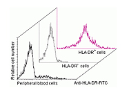
We frequently use Anti-HLA-DR MicroBeads for positive selection of antigen presenting cells (APCs) from peripheral blood mononuclear cell (PBMC) suspensions, either directly from fresh preparations, or after stimulation with different antigens. After separation, we isolate RNA from APCs. The protocol can be divided into two major steps, magnetic labelling and magnetic separation. Both parts are easy to handle and together take about 50-60 minutes. There is virtually no loss of cells when separating APCs from fresh PBMCs. The yield is about 17-22% of the full PBMC count, which is the expected fraction of APCs in PBMC suspensions. Purity repeatedly reaches more than 95% as determined by fluorescence activated cell sorting. However, if we stimulate PBMCs with antigen prior to positive selection of HLA-DR bearing cells, we commonly only gain about 30-70% of the expected APC count. Purity ranges between 70-80%. These observations might, at least in part, be explained by cell lysis during the incubation period. Moreover, down-regulation of HLA-DR on the cell surface cannot be excluded. To reach higher purities, it is possible to reload the eluted APCs on a new freshly prepared column.
PhD Student
Institute f. Med. Microbiology & Hygiene
University of Regensburg