In a paper published recently in Biomaterials, researchers from Okayama University described their new method to more effectively recreate pancreatic cancer tissue in vitro. Existing experimental models have not yet been able to fully replicate the extent of fibrosis in human tissue, they reported.
Using a three-dimensional (3D) cell culture technique, the team devised a method to mix pancreatic cancer cells together with fibrotic components to generate tissues that resemble human pancreatic cancer.
According to senior author Professor Mitsunobu R. Kano, "No cancer is an island entire of itself. In pancreatic cancer, fibrotic tissue often occupies much more space than the cancer cells themselves. While numerous 3D models of pancreatic cancer have been reported, ours is the first to allow the tuning of the amount of fibrosis in the model."
The team set about creating the novel 3D model in order to gain a better understanding of the fibrotic mechanisms in pancreatic cancer and how the thick fibrotic tissue obstructs the penetration of drugs into the tumor and limits anti-tumor immunity.
Search Antibodies Search Now Use our Antibody Search Tool to find the right antibody for your research. Filter
by Type, Application, Reactivity, Host, Clonality, Conjugate/Tag, and Isotype.
Since existing in vitro models failed to replicate the fibrotic components that are seen in pancreatic cancer, the team reasoned that it was important to establish relevant pancreatic cancer models that comprise not only cancer cells but also cells involved in fibrosis. They also found that the area occupied by fibrosis in human pancreatic cancer tissues ranges from 40 to 80% among patients, so the team needed to make sure it could recreate tissue with any given amount of fibrosis."
Fibrosis consists mainly of fibroblasts and other extracellular matrix components secreted by fibroblasts. Considering this, the research team came up with a simple strategy: mix and culture pancreatic cancer cells and fibroblasts at various ratios. "The strategy seemed too simple to work," lead author Hiroyoshi Y. Tanaka explained.
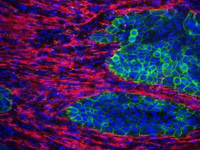
The team successfully showed that by varying the ratio of pancreatic cancer cells seeded against a fixed number of fibroblasts, they could create pancreatic cancer tissues in vitro with different amounts of fibrosis. Importantly, they showed that the area occupied by fibrosis in these tissues can be experimentally tuned to match the amount of fibrosis in the clinically observed range. The team also went on to use the model to successfully understand the molecular mechanisms by which a particular phenotype of fibroblasts, characteristic to pancreatic cancer, occurs.
The scientists are optimistic that this novel model can complement other well-established models of pancreatic cancer, especially with regard to fibrosis. "The great thing about our model is that it is fairly simple to create," concluded Prof Kano. "We believe it will be useful in mechanistic studies as well as in screening for drugs that target fibrosis. This is just the beginning, but we hope to make a real difference in the treatment of pancreatic cancer."
Image: Immunofluorescence staining image of the 3D fibrotic tissue. Cancer cells (green) exist embedded within a densely fibrotic extracellular matrix (red). Cell nuclei are stained in blue. Image courtesy of Mitsunobu R. Kano.