Glaucoma, the leading cause of irreversible blindness globally, affects over 60 million people, which is predicted to double by 2040 as the global population ages. Loss of sight in glaucoma is caused by the death of cells in the retina at the back of the eye. But in a study published yesterday in Expert Review of Molecular Diagnostics, researchers share a new technology that can detect glaucoma progression 18 months earlier than the current gold standard method.
“We have developed a quick, automated, and highly sensitive way to identify which people with glaucoma are at risk of rapid progression to blindness,” says senior author Francesca Cordeiro of Imperial College London.
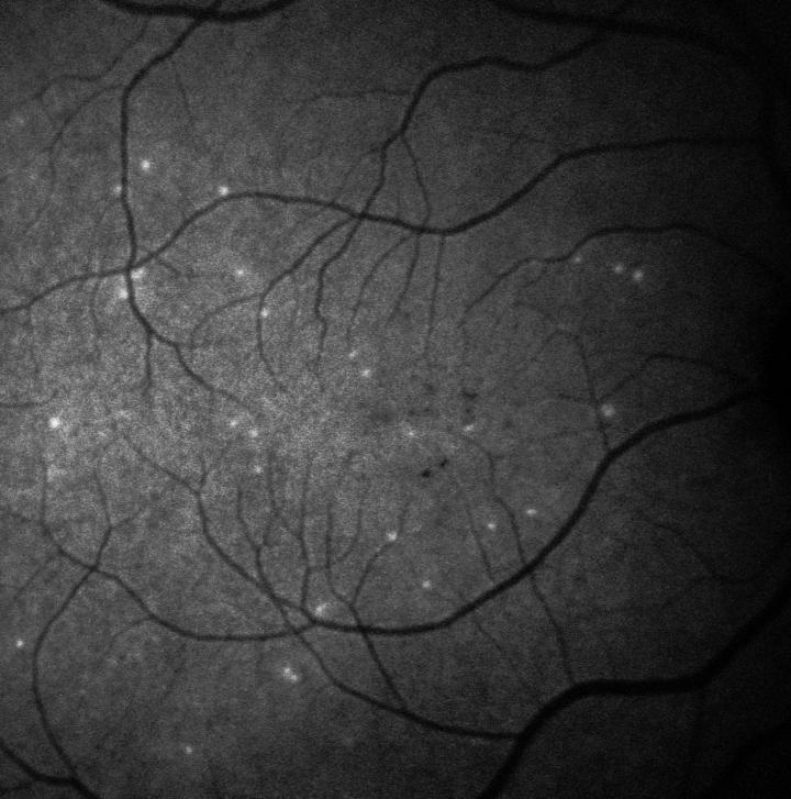
The test, called DARC (Detection of Apoptosing Retinal Cells), involves injecting a fluorescent dye into the bloodstream that attaches to retinal cells, illuminating those that are in the process of apoptosis. The damaged cells appear bright white when viewed in eye examinations. Additionally, the researchers incorporated an AI algorithm into their method for objective DARC count analysis.
Search Antibodies Search Now Use our Antibody Search Tool to find the right antibody for your research. Filter
by Type, Application, Reactivity, Host, Clonality, Conjugate/Tag, and Isotype.
In the Phase II clinical trial of DARC, the AI was used to assess 60 of the study participants (20 with glaucoma and 40 healthy control subjects). The AI was initially trained by analyzing the retinal scans (after injection of the dye) of the healthy control subjects, after which it was then tested on the glaucoma patients. The researchers followed up 18 months later to see whether the patients’ eye health had deteriorated.
Using the AI technology, the researchers were able to accurately predict progressive glaucomatous damage 18 months earlier than they can with the current gold standard OCT retinal imaging technology, as every patient with a DARC count over a certain threshold was found to have progressive glaucoma at follow-up.
“These results are very promising as they show DARC could be used as a biomarker when combined with the AI-aided algorithm,” Cordeiro says. “What is really exciting, and actually unusual when looking at biological markers, is that there was a clear DARC count threshold above which all glaucoma eyes went on to progress.”
The team is also applying the test to rapidly detect cell damage caused by numerous conditions other than glaucoma, such as other neurodegenerative conditions that involve the loss of nerve cells, including age-related macular degeneration, multiple sclerosis, and dementia.
Image: A patient's retina showing hyperfluorescent signals—each white spot is a single 'sick' retinal nerve cell. Imagery supported by DARC technology used in the Phase II clinical trial. Image courtesy of UCL/Western Eye Hospital.