Biological robots, or biobots, draw inspiration from natural systems to mimic the motions of organisms, such as swimming or jumping. Improvements to biobots to better replicate complex motor behaviors can lead to exciting biorobotic engineering applications to help solve real world challenges. However, this requires the creation of biohybrid robots—biobots made up of both organic and artificial materials—which is a challenge.
Researchers from the University of Illinois at Urbana–Champaign combined an intact rat spinal cord with a tissue-engineered 3D muscle system. They describe the novel biohybrid system in a report published today in APL Bioengineering.
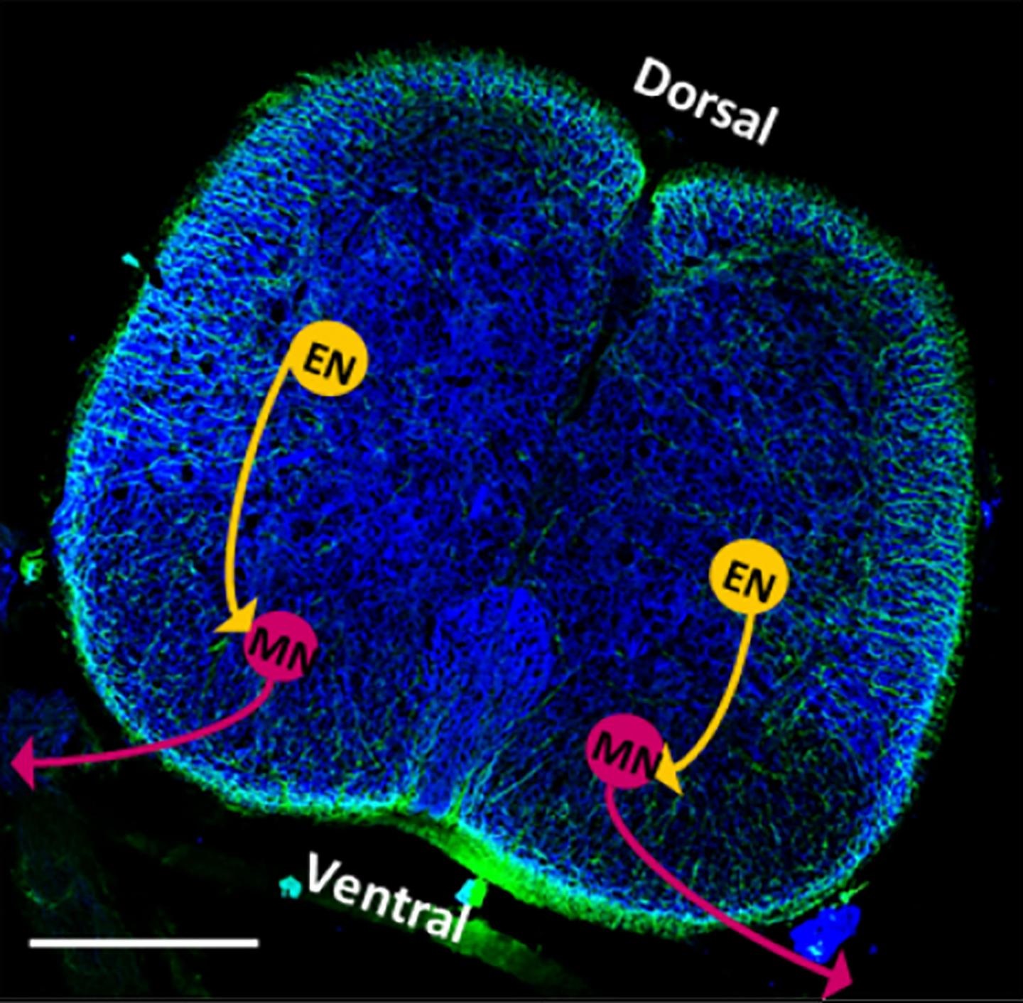
After culturing the system for seven days, the researchers found that the motor neurons from the spinal cord begin to produce electrical activity that causes contraction in the artificial muscles, mirroring the behavior of the peripheral nervous system.
Search Antibodies Search Now Use our Antibody Search Tool to find the right antibody for your research. Filter
by Type, Application, Reactivity, Host, Clonality, Conjugate/Tag, and Isotype.
“When we looked more deeply at how the neuron–muscle interface developed, we were very excited to observe many similarities between our tissue-engineered spinobot and in vivo development,” says first author Collin Kaufman. According to Kaufman, this result indicates that the explanted spinal cord is a viable mechanism for controlling muscular behaviors, even when removed from its natural environment.
The researchers further tested this by varying the concentration of neurotransmitters in the system. When additional neurotransmitters are present, the contractions are more patterned and consistent, and when they are blocked, the twitching decreases.
Because studying the peripheral nervous system can be so difficult, the ability to observe it externally—as demonstrated in the present study—can lead to great strides in medicine. One potential example is amyotrophic lateral sclerosis, in which the death of neurons results in the eventual loss of motor function. By developing an external peripheral nervous system, researchers can study ALS with ease of access to the impacted components in real time.
“The next steps to studying such a disease are surprisingly close,” Kaufman says. “By replacing the muscle, the spinal cord, or any combination of the two tissues with an ALS mutant model, researchers would be able to study how diseased neurons interact with nearby muscles.”
Image: A cross-section of a rat's spinal cord displays the presence of neurons and neuronal stimulation. Image courtesy of Collin D. Kaufman.