The 2014 Nobel Prize in Chemistry was awarded for the development of super-resolution fluorescence microscopy technology that afforded microscopists the first molecular view inside cells, a capability that has provided new molecular perspectives on complex biological systems and processes. In a study published Friday in Science Advances, UNSW researchers pushed our resolution capabilities even further.
Our current super-resolution techniques allow for the observation of individual molecules; however, interactions between these molecules occur at a scale at least four times smaller than that resolved by existing single-molecule microscopes.
“The reason why the localization precision of single-molecule microscopes is around 20–30 nanometers normally is because the microscope actually moves while we’re detecting that signal,” explains senior author Katharina Gaus. “This leads to an uncertainty. With the existing super-resolution instruments, we can’t tell whether or not one protein is bound to another protein because the distance between them is shorter than the uncertainty of their positions.”
Search Antibodies Search Now Use our Antibody Search Tool to find the right antibody for your research. Filter
by Type, Application, Reactivity, Host, Clonality, Conjugate/Tag, and Isotype.
To circumvent this problem, the team built autonomous feedback loops inside a single-molecule microscope that detects and realigns the optical path and stage.
“It doesn’t matter what you do to this microscope, it basically finds its way back with precision under a nanometer,” Gaus says. “It’s a smart microscope. It does all the things that an operator or a service engineer needs to do, and it does that 12 times per second.”
The feedback system designed by the UNSW team is compatible with existing microscopes and affords maximum flexibility for sample preparation. “We just built a microscope within a microscope, and all it does is align the main microscope,” Gaus explains.
The researchers used their microscope to perform direct distance measurements between signaling proteins in T cells. A popular hypothesis in cellular immunology is that these immune cells remain in a resting state when the T cell receptor is next to another molecule that acts as a brake. With their high-precision microscope, the researchers showed that these two signaling molecules are in fact further separated from each other in activated T cells, releasing the brake and switching on T cell receptor signaling.
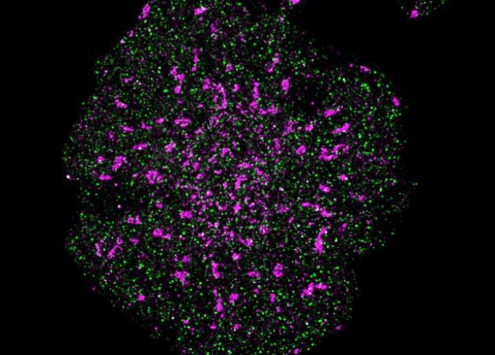
“Conventional microscopy techniques would not be able to accurately measure such a small change as the distance between these signaling molecules in resting T cells and in activated T cells only differed by 4–7 nanometers,” Gaus says. “This also shows how sensitive these signaling machineries are to spatial segregation. In order to identify regulatory processes like these, we need to perform precise distance measurements, and that is what this microscope enables.”
Image: A T cell with precise localization of T cell receptors (pink) and CD45 phosphatase (green). Image courtesy of Single Molecule Science, UNSW Sydney.