It is increasingly recognized that the pH directly surrounding a cell is an important indicator of cellular health; in fact, it seems to play a role in cancer progression. However, techniques to measure this pH have so far remained limited in terms of their sensitivity, their spatial resolution, and their response time to pH changes. In a study published today in Nature Communications, researchers describe a nanopipette pH biosensor that is sensitive to changes in pH of less than 0.01 units with a response time of 2 ms and 50 nm spatial resolution.
The researchers originally designed the sensor as a nanopipette ionic field effect transistor in which gates control the flow of ions in the nanopipette instead of electrons. However, while this tackled issues around pH sensitivity and spatial resolution, the device readings still took a few seconds to respond to pH changes due to ionic Coulomb blockade effects hampering the diffusion rate of ions.
Search Antibodies Search Now Use our Antibody Search Tool to find the right antibody for your research. Filter
by Type, Application, Reactivity, Host, Clonality, Conjugate/Tag, and Isotype.
The solution the researchers now propose is to incorporate a zwitterionic membrane to enable faster responses. By using a twin barrel nanopipette with the membrane in just one of the barrels, the researchers were able to use the other barrel as a scanning ionic conductance microscope (SICM) for simultaneous topological measurements.
The team tested the device on live cancer cells and showed how the device could pick up on increases in extracellular pH from invasive phenotypes of breast cancer cells that had been deprived of estrogen. They could also detect pH changes from algae exposed to sunlight, caused by the uptake of inorganic carbon in photosynthesis, as well as identifying heterogeneities in aggressive melanoma cells from high-resolution pH maps.
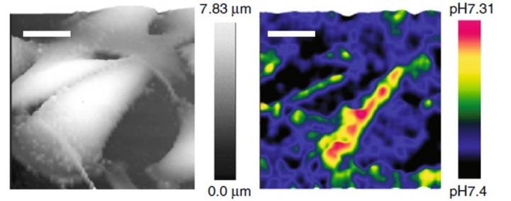
The new tool allows for real-time, feedback-controlled, dynamic 3D mapping of extracellular pH, and it can detect multiple types of cancer cells at a subcellular resolution without requiring any labels. According to the researchers, this method could be beneficial for the diagnosis and prognosis of cancer and in evaluating therapies targeted to extracellular pH.
Image: High-resolution 3D pHe mapping of living melanoma cells with feedback-controlled double-barrel SICM-pH nanoprobe. The 3D SICM topographical images (left column) and 3D pHe distributions (right column) of low-buffered living melanoma A375M obtained simultaneously by a single SICM scanning, which demonstrated a highly variegated distribution pattern of pHe. Scale bars represent 20 μm. Image courtesy of Nature Communications and Kanazawa University.