George Mason University researchers have discovered the exact location where two proteins responsible for hiding cancer cells from the immune system bind. This discovery provides a novel approach to developing new cancer immunotherapy medicines that can be administered as a pill, compared to existing intravenous therapeutics. The findings were published recently in the Journal of Biological Chemistry.
According to first author Amanda Haymond, the discovery was made possible by an in-house developed protein-painting technology. The process starts with two or more proteins that, when bound together, drive disease. The scientists use small-molecule dyes to paint the bound proteins and then they chemically separate them. Finally, they use a mass spectrometer to identify the unpainted regions, which is where the proteins touch.
Search Antibodies Search Now Use our Antibody Search Tool to find the right antibody for your research. Filter
by Type, Application, Reactivity, Host, Clonality, Conjugate/Tag, and Isotype.
Current technologies in early-phase drug discovery, such as crystallography, are often complicated, costly, and time-consuming. The protein-painting technology specifically identifies protein–protein touchpoints, highlighting an ideal location and recipe to follow for drug development. The recipe, along with the fact that the technology allows for rapid performance testing of the drug, means that results can be produced in days rather than years.
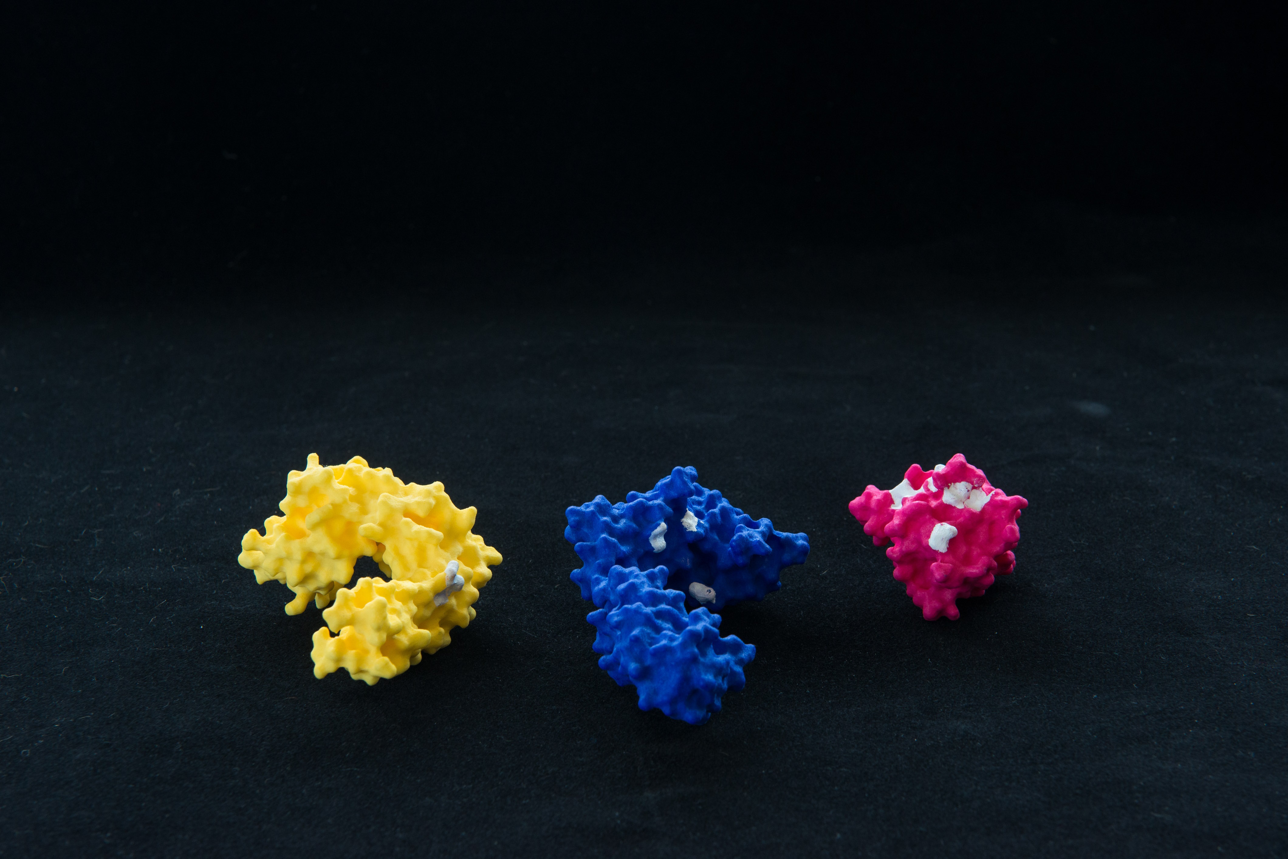
In the new article, the team describes how they enhanced their technology, reporting the development and optimization of a novel protein dye that has been successfully tested on clinically relevant protein complexes PD-1 and PD-L1. The publication also unveils new findings that chemically decipher the way that dyes interact with proteins, which has been a mystery to scientists for decades.
“The secret to using the protein-painting technique is having the perfect dye molecule with just the right structure to bind tightly onto proteins,” says Haymond.
Image: These 3D models of a receptor, co-receptor, and ligand demonstrate how the protein painting technology works. Bound proteins in their native form are coated with small molecule dyes. The regions that aren’t painted when the proteins are bound—designated white—can be detected by mass spectrometry and inform rational drug development. Image courtesy of Evan Cantwell.