Researchers at MIT and the Broad Institute of Harvard and MIT have devised a new way to rapidly image synaptic proteins at high resolution. Using fluorescent nucleic acid probes, they can label and image an unlimited number of different proteins. They demonstrated the technique, called PRISM, in a new study in which they imaged 12 proteins in cellular samples containing thousands of synapses.
"Multiplexed imaging is important because there's so much variability between synapses and cells, even within the same brain," explains Mark Bathe, senior author of the study that was published today in Nature Communications. "You really need to look simultaneously at proteins in the sample to understand what subpopulations of different synapses look like, discover new types of synapses, and understand how genetic variations impact them."
Synaptic proteins have a variety of functions. Many of them help to form synaptic scaffolds, which are involved in secreting neurotransmitters and processing incoming signals. While synapses contain hundreds of these proteins, conventional fluorescence microscopy is limited to imaging at most four proteins at a time. To boost that number, the MIT team developed a new technique based on an existing method called DNA PAINT. Using this method, researchers label proteins or other molecules of interest with a DNA-antibody probe. Then, they image each protein by delivering a fluorescent DNA "oligo" that binds to the DNA-antibody probes.
Search Antibodies Search Now Use our Antibody Search Tool to find the right antibody for your research. Filter
by Type, Application, Reactivity, Host, Clonality, Conjugate/Tag, and Isotype.
The DNA strands have an inherently low affinity for each other, so they bind and unbind periodically, creating a blinking fluorescence can be imaged using super-resolution microscopy. However, imaging each protein takes about half an hour, making it impractical for imaging many proteins in a large sample.
Bathe and his colleagues set out to create a faster method that would allow them to analyze a huge number of samples in a short period of time. To achieve that, they altered the DNA-dye imaging probe so that it would bind more tightly to the DNA-antibody, using locked nucleic acids. This gives a much brighter signal, so the imaging can be done more quickly, but at slightly lower resolution.
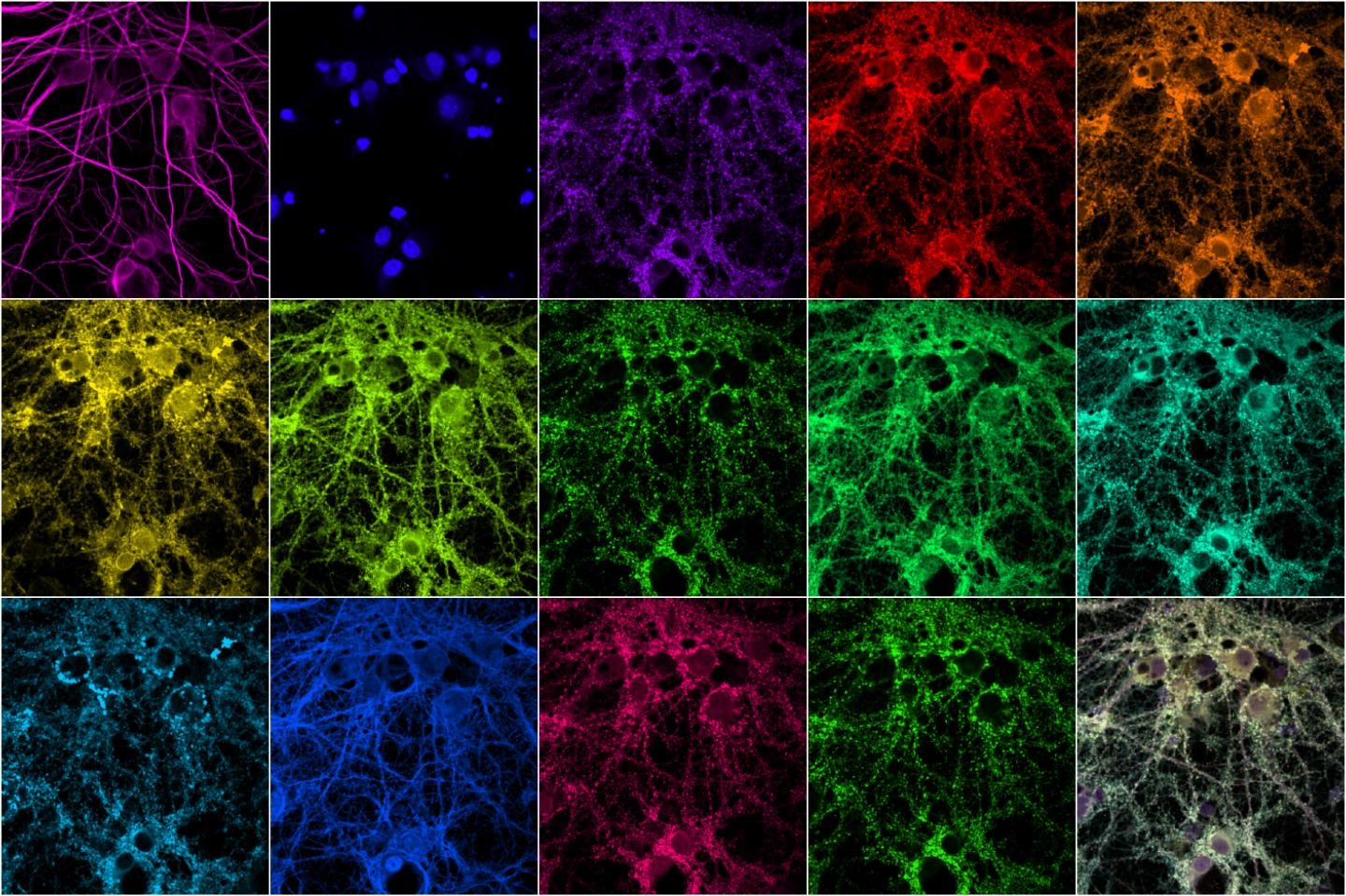
Image: MIT engineers have developed a technique that allows them to rapidly image many different proteins within a synapse. At bottom right is a composite of the other images. Image courtesy of Syuan-Ming Guo and Li Li.