Small-cell clones in proliferating epithelia—tissues that line all body surfaces—organize very differently than their normal-sized counterparts, according to a study from the Stowers Institute for Medical Research published today in Developmental Cell.
"A common feature of many cancer types is the pleomorphic nature—or variability in size and shape—of cells in a tumor," says first author Subramanian P. Ramanathan, Ph.D. "What's not exactly known is whether differential cell size is the driver for, or the result of, cancer progression."
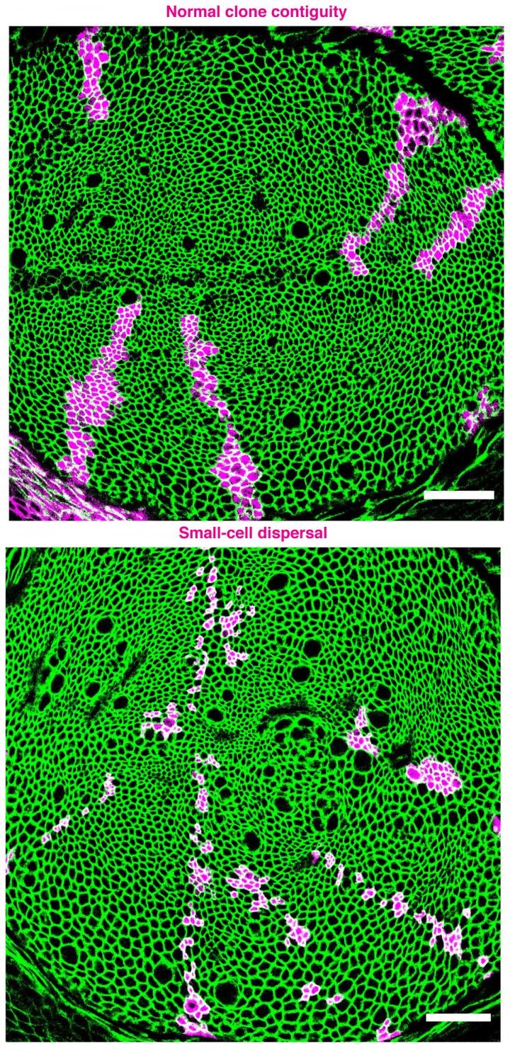
Cells in an epithelial sheet are normally connected by Velcro-like structures called adhesive junctions and have a near-uniform size distribution. As a result, cells related by descent tend to stick together in mosaic patches referred to as cell clones. In the fruit fly Drosophila melanogaster, epithelial cell clones that have smaller cells can lose contact with each other and disperse among their neighbors.
The striking dispersal of small cells carrying mutations in a gene called Tor was first observed over a decade ago by senior author Matthew Gibson, Ph.D. when he was a postdoctoral researcher. "I puzzled over it, but just couldn't make sense of why the mutant cells dispersed within the cell layer," says Gibson. Not until 2015, when Ramanathan joined the lab, did they begin to piece together the science behind this observation.
Search Antibodies Search Now Use our Antibody Search Tool to find the right antibody for your research. Filter
by Type, Application, Reactivity, Host, Clonality, Conjugate/Tag, and Isotype.
"We initially thought we were going to be studying cell division and its relationship to junction formation," says Gibson. "But based on Subramanian's experiments, we determined that the Tor cells divide and make a junction that later becomes unstable. He was able to rule out cytoskeletal or biophysical explanations, and that led to a mathematical treatment of the problem."
"The eureka moment was the soap bubbles experiment," says Ramanathan, citing D'Arcy Wentworth Thompson's, On Growth and Form. "About a hundred years ago, Thompson documented the similarity between how soap foam and biological tissue organize. Soap cells grow and shrink in an extremely predictable fashion. We found that the junctions shared by smaller soap cells frequently collapsed separating the small cells. This was strikingly similar to what we saw in epithelial tissue," says Ramanathan.
Ramanathan reasoned that the Tor cell dispersal could be a geometrical consequence. To explore it in the purely in silico framework, they reached out to co-author Matej Krajnc, Ph.D., from Princeton University, who has experience with these types of models. "He was excited to modulate cell size, to see if we could see similar patterns emerging in silico. And that is indeed what we saw."
One of the most astounding feats of developmental biology is the transformation of an epithelial sheet into a functionally specialized three-dimensional structure. To do so, epithelial systems rely on uniformity among cells. Developmental processes shape and sculpt epithelia by creating region-specific properties that are classically understood to be driven by molecular pathways.
"In the human body, which is primarily made up of epithelial cell layers, the vast majority of mutations we accumulate are not germline, but rather somatic, and therefore clonal in nature," says Gibson. "In a pathogenesis context, because mutations create clonal effects, we don't have a framework for thinking about how clones of cells behave when they're genetically distinct from the surrounding wild type cells, but it's extremely important."
Image: Normal cell clone arrangement in which they clump together (top, magenta) and mutant clones comprised of smaller cells aberrantly dispersed within epithelial tissue (bottom). Image courtesy of Gibson Lab, Stowers Institute for Medical Research.