Langerhans cell histiocytosis (LCH) is an unusual disease. Often classified as a cancer because of uncontrolled cell growth in different parts of the body, it also has features of an autoimmune disease, as LCH lesions attract immune cells and show characteristic tissue inflammation. In a study published yesterday in Cancer Discovery, researchers analyzed LCH cell types and discovered a developmental hierarchy in these cells.
LCH is clinically variable and often difficult to diagnose. Skin involvement in babies with LCH can look like a nappy rash, whereas bone involvement can be mistaken as sarcoma in an x-ray picture. In its most aggressive form, LCH can present as a leukemia-like disease and lead to organ failure. These diverse manifestations and the enormous clinical heterogeneity of LCH continue to puzzle medical doctors and scientists around the world.
Search Antibodies Search Now Use our Antibody Search Tool to find the right antibody for your research. Filter
by Type, Application, Reactivity, Host, Clonality, Conjugate/Tag, and Isotype.
Studying LCH lesions under the microscope, senior author Caroline Hutter of St. Anna Children’s Hospital observed striking heterogeneity among LCH cells. Her aim was to answer two fundamental questions: What are the mechanisms behind LCH, and how can we improve treatment of children affected by this disease?
Utilizing state-of-the-art technology, the team analyzed LCH lesions for their molecular composition at single-cell resolution. The team analyzed the molecular profiles of LCH lesions and developed a comprehensive map of cellular heterogeneity in LCH.
The researchers identified multiple LCH cell subtypes. One of these subtypes comprised actively dividing cells, which appear to give rise to the other LCH cell subtypes. In further experiments, the team unraveled the molecular pathways that are active in different branches of this unexpected developmental hierarchy, which corroborated an interplay of developmental, immunological, and oncogenic mechanisms in LCH.
The study is a significant step toward understanding this enigmatic disease. In the future, these findings may lead to better methods of distinguishing severe from less severe disease cases, and they may even open up new treatment possibilities.
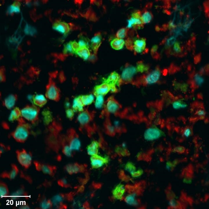
Image: These are LCH cells from a tumor biopsy, imaged with a confocal microscope. The green staining identifies the actively proliferating cells. Image courtesy of CCRI, Vienna.