The disability burden for people with multiple sclerosis (MS) depends largely on the form of the disease that they have: The relapsing/remitting form of the disease is associated with periods of clinical remission, while the progressive form is associated with continued neurological deterioration without clinical remission. Effective therapies exist for managing relapsing/remitting MS, but treatment for progressive MS has proved more challenging. But a paper published today in Brain has identified potential mechanisms that may inform the development of therapies to effectively manage progressive MS.
“Because the brain is bathed by the cerebrospinal fluid (CSF), we asked whether treating cultured neurons with the CSF from MS patients with a relapsing/remitting or a progressive disease course would possibly elicit different effects on neuronal mitochondrial function,” says senior author Patrizia Casaccia of the City University of New York. “We detected dramatic differences in the shape of the neuronal mitochondria and their ability to produce energy. Only exposure to the CSF from progressive MS patients caused neuronal mitochondria to fuse and elongate while rendering them unable to produce energy.”
Search Antibodies Search Now Use our Antibody Search Tool to find the right antibody for your research. Filter
by Type, Application, Reactivity, Host, Clonality, Conjugate/Tag, and Isotype.
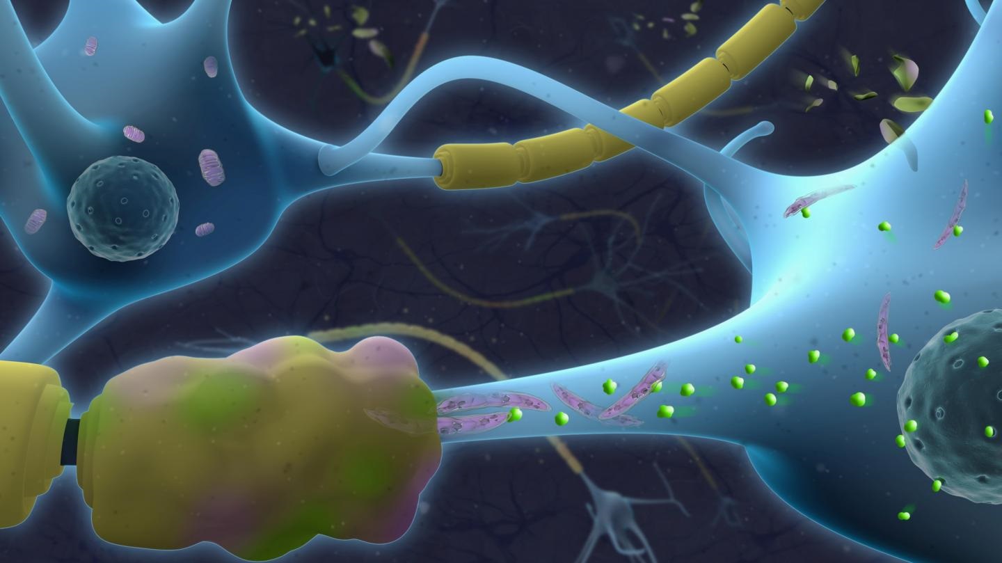
CSF samples were acquired from 15 patients with relapsing/remitting MS and 29 with progressive MS. These samples were extensively characterized—both functionally and metabolically. Video recordings of live, cultured rat neurons revealed that mitochondria exposed to CSF samples from progressive MS patients tended to be elongated, while mitochondria exposed to CSF from relapsing/remitting MS patients were not. They also found through lipid profiling that CSF from progressive MS patients has increased levels of ceramides.
“When we exposed cultured neurons to ceramides, we elicited the same changes caused by exposure to CSF from progressive MS patients, and we further discovered that ceramides induced neuronal damage by acting on two cellular mechanisms,” says first author Maureen Wentling, also of CUNY. “On one end, ceramides impaired the ability of neurons to make energy by directly damaging the mitochondria. On the other end, they also forced neurons to more rapidly uptake glucose in an attempt to provide energy for the cell.”
The researchers were able to prevent the neurotoxic effect of CSF on cultured neurons by supplementing glucose. Supplementation isn’t a sustainable approach in the diseased brain, however, so a different approach will ultimately be needed for developing therapies that improve mitochondrial function in patients with progressive MS.
Image: The top portion of this graphic features Neurons (blue) with axons wrapped by normal myelin (yellow). The lower portion of this graphic illustrates neurons with pathological axons wrapped by damaged myelin (yellow and pink). The elongated mitochondria (purple) in the lower portion are dysfunctional and characterized by accumulation of toxic ceramides (green). Image courtesy of Jeremy Weichsel at Biovisioning.