A new type of imaging that can see what’s happening in cells at the genomic level has been developed by Howard Hughes Medical Institute scientists. DNA microscopy uses DNA bar codes to help pinpoint molecules' relative positions within a sample. According to the team, it's an entirely new category of microscopy, “it's not just a new technique, it's a way of doing things that we haven't ever considered doing before."
With DNA microscopy, scientists can build a picture of cells and simultaneously amass enormous amounts of genomic information, reports Joshua Weinstein, first author on the paper published today in Cell. "This gives us another layer of biology that we haven't been able to see."
Until now, microscopy fit into two main categories. Optical imaging can offer intricate portraits of subcellular structure and action. Dissection-based microscopy can give scientists genetic information.
Search Antibodies Search Now Use our Antibody Search Tool to find the right antibody for your research. Filter
by Type, Application, Reactivity, Host, Clonality, Conjugate/Tag, and Isotype.
Weinstein and his colleagues wanted to create a way to do it all in one shot—to take a snapshot of a cell's position and spell out the specific genetic sequences driving it. That combo is important for scientists studying genetically diverse sets of cells. The immune system is a perfect example, Weinstein says. Immune cell genes can vary down to a single letter of DNA. Each variation can trigger a dramatic shift in the type of antibodies a cell produces. Where that cell is located within a tissue can alter antibody production, too. If you focus on just one or the other, "you're only getting part of the picture," he says.
The new system doesn't require an expensive microscope or a lot of fancy equipment. First, scientists take cells grown in the lab and fix them into position in a reaction chamber. Then, they add an assortment of DNA bar codes. These stick to RNA molecules, giving each a unique tag. Next, the team uses a chemical reaction to make more and more copies of each tagged molecule—a growing pile that expands out from each molecule's original location.
Eventually, the tagged molecules collide with other tagged molecules, forcing them to link together in pairs. Molecules located close to one another will be more likely to collide, generating more DNA pairs. Molecules further apart will generate fewer pairs.
A DNA-sequencing machine spells out the letters of every molecule within the sample, which takes up to 30 hours. An algorithm the team created then decodes the data—which, in the paper, represents roughly 50 million DNA letters of genetic sequences from each original specimen—and converts the raw data to images.
"You're basically able to reconstruct exactly what you see under a light microscope," Weinstein says. The two methods are complementary, he adds. Light microscopy can see molecules well even when they're sparse within a sample, and DNA microscopy excels when molecules are dense—even piled up on top of one another.
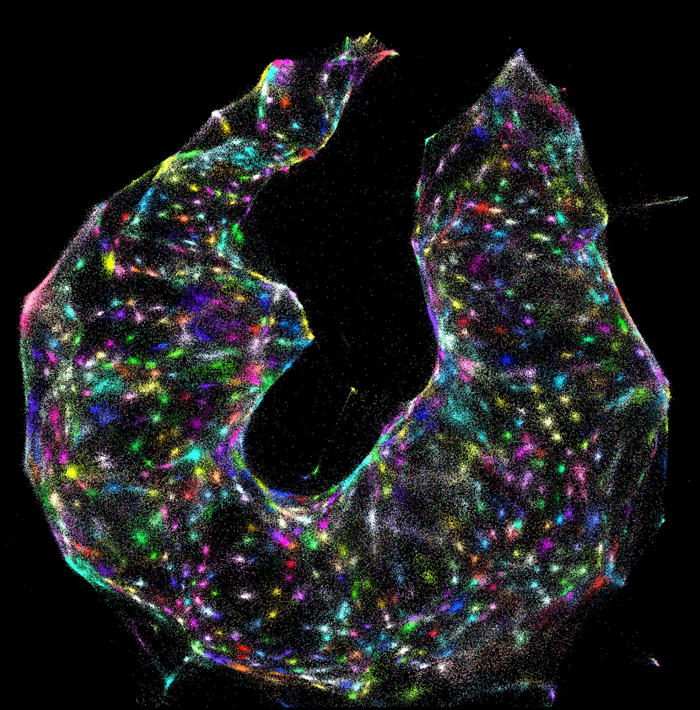
Image: Using DNA microscopy, scientists can identify different cells (colored dots) within a sample—with no prior knowledge of what the sample looks like.