The diagnosis and treatment of cancer is a challenge for physicians, and part of what makes it so challenging is tumor heterogeneity. But researchers have now shown that harmless purple bacteria of the genus Rhodobacter can be used to visualize heterogeneity in tumors. With the aid of optoacoustic imaging, the researchers used these microorganisms to visualize immune cells called macrophages that play a role in tumor development. The findings were published today in Nature Communications.
Many cancers form solid tumors. Inside the tumors, there are major differences at the cellular and molecular level, including the localization and activity of macrophages. Although macrophages are essential for a healthy immune system, they also play a key role in tumor development. With the aid of photosynthetic bacteria, the researches have developed new optoacoustic techniques that indicate where such macrophages are present and active.
“We were able to demonstrate that bacteria of the genus Rhodobacter, which are harmless to humans, are suitable as indirect markers of macrophage presence and activity,” says senior author Andre C. Stiel of Helmholtz Zentrum München.
Search Antibodies Search Now Use our Antibody Search Tool to find the right antibody for your research. Filter
by Type, Application, Reactivity, Host, Clonality, Conjugate/Tag, and Isotype.
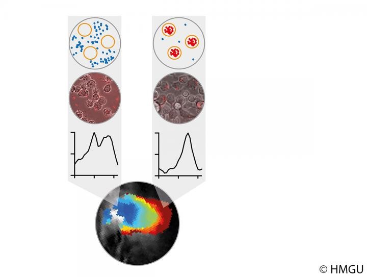
Rhodobacter bacteria produce large quantities of the photosynthetic pigment bacteriochlorophyll a. This pigment enabled the researchers to detect bacteria in a tumor by means of multispectral optoacoustic tomography (MSOT).
How does the principle work? Macrophages engulf bacteria as part of their natural scavenging activity. This alters the surroundings of the bacteria, their absorption of electromagnetic radiation and, as a result, also the optoacoustic signal. Rhodobacter bacteria thus act like sensors for scientists, providing them with information about the presence and activity of macrophages.
“In further steps, these bacteria will enable novel approaches to non-invasive technologies and so open up entirely new possibilities for innovative diagnostic and therapeutic procedures,” says coauthor Thomas Drepper of Heinrich Heine University Duesseldorf.
In the future, bacteria may be able to reveal the location of a tumor and also detect increased macrophage activity. Depending on their localization, the macrophages could provide information about unwelcome inflammations or the desired response to immunotherapies and could ultimately be used to improve treatment strategies.
Image: Change of optoacoustic signals of purple bacteria located outside (blue) and inside (red) of macrophages visualized via schematic (first row), microscopic (second row) and MSOT analysis (bottom). The change of MSOT spectra (third row) can be used to differentiate between Rhodobacter cells located inside and outside of macrophages and hence macrophage localization and activity. Image courtesy of Helmholtz Zentrum München.