Spontaneous recovery from spinal cord injury is almost unheard of in humans and other mammals, but many vertebrates fare better. The eel-like lamprey, for instance, can fully regenerate its spinal cord even after it has been severed. Three months after injury, the lamprey is swimming, burrowing, and flipping again, as if nothing had happened.
In a study published today in PLOS ONE, scientists report that lampreys recover and regenerate just as well after a second complete spinal cord injury at the same location. The study opens up a new path for identifying pro-regenerative molecules and potential therapeutic targets for human spinal cord injury.
“We’ve determined that central nervous system (CNS) regeneration in lampreys is resilient and robust after multiple injuries,” says senior author Jennifer Morgan of the Marine Biological Laboratory. “The regeneration is nearly identical to the first time, both anatomically and functionally.”
Search Antibodies Search Now Use our Antibody Search Tool to find the right antibody for your research. Filter
by Type, Application, Reactivity, Host, Clonality, Conjugate/Tag, and Isotype.
Morgan’s lab has been focusing on the descending neurons, which originate in the brain and send motor signals down to the spinal cord. Some of these descending neurons regenerate after CNS injury in lamprey, while others die.
“We are beginning to isolate individual descending neurons and look at their transcriptional profiles (gene activity) to see if we can determine what makes some of them better at regenerating than others,” Morgan says. “The ‘good’ regenerators, for example, may express molecules that are known to promote growth during development. That’s one hypothesis.”
Observing how the descending neurons respond to a second CNS injury can help the team tease out the factors required for repeated, resilient regeneration, which could have implications for designing better strategies for treatments aimed at promoting CNS re-growth after injury or disease.
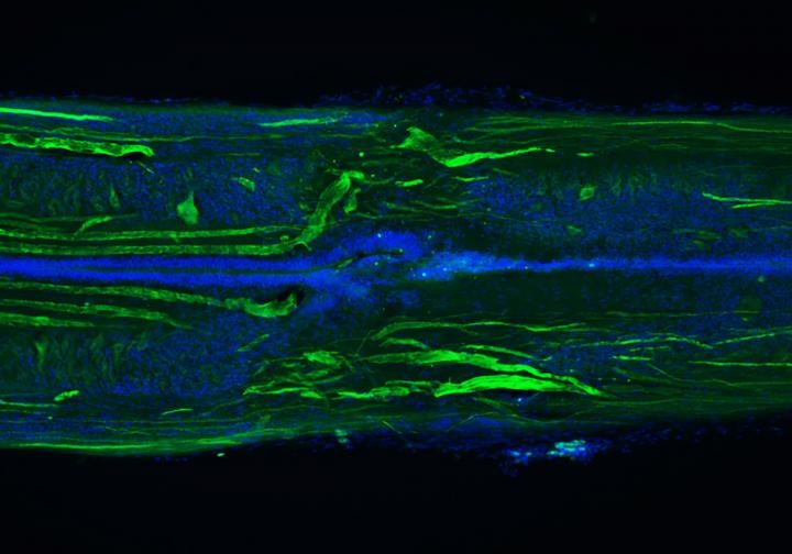
Image: Longitudinal section of a lamprey spinal cord at 11 weeks post-injury, showing many regenerated axons (green) and a repaired central canal (blue tubelike structure). The original lesion site is in the center of the image. Image courtesy of S. Allen and J. Morgan.