Super-resolution microscopy allows scientists to observe cells and their interior structures and organelles in a way never before possible. But it does have some drawbacks, including that it typically can only use one or two fluorescent colors, which makes it difficult to observe different proteins and decipher the complex architecture and underlying assembly mechanisms of the cell's interior structures. In addition, overcoming the noise inherent to super-resolution methods and fluorescent labeling in order to achieve the full resolution potential is challenging.
Scientists from EPFL report that they have now solved both problems by developing a new method to analyze and reconstruct super-resolution images and re-align them in a way that multiple proteins can be placed within a single 3D volume. The method, described in a Nature Methods paper published today, works with images taken with large field-of-view super-resolution microscopy, with each image containing hundreds of two-dimensional projections of a labeled structure in parallel.
Each 2D view represents a slightly different orientation of the structure, so that with a dataset of thousands of views, the method can computationally reconstruct and align the 2D images into a 3D volume. By combining information from a large number of single images, the noise is reduced and the effective resolution of the 3D reconstruction is enhanced.
With the help of Pierre Gönczy's lab at EPFL, the researchers tested the method on human centriole complexes. Using the new multicolor super-resolution reconstruction method, the researchers were able to uncover the 3D architecture of four proteins critical for centriolar assembly during organelle biogenesis.
The new approach allows for unlimited multiplexing capabilities. "With this method, if the proteins in the structure can be labeled, there is no limit to the number of colors in the 3D reconstruction," says Suliana Manley, senior author. "Plus, the reconstruction is independent of the super-resolution method used, so we expect this analysis method and software to be of broad interest."
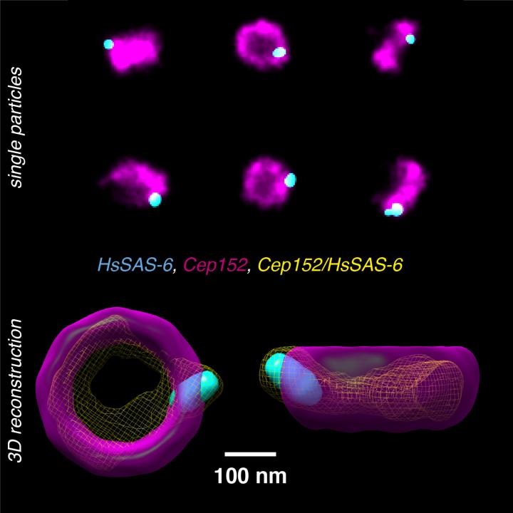
Image: Human centrioles labeled with antibodies against two proteins (Cep152, HsSAS-6) and imaged using super-resolution microscopy. From many individual particles showing projections of the centriole complex in various orientations (upper panel), by using a fused intermediate (yellow, lower panel), the newly developed method allows now to reconstruct a multicolor 3D model (lower panel). Image courtesy of Christian Sieben/EPFL.