A new cell culture platform allows researchers to observe never-before-seen behaviors of live cancer cells under the microscope, according to researchers at Hokkaido University. They reported in a Scientific Reports paper today that their easy-to-produce platform features micro-scale attachment sites for cancer cells that enable observation of behaviors highly relevant to cancer's clinical properties.
"Cancer studies so far either use cell cultures in which cancer cells don't necessarily behave naturally, or tissue samples that don't allow live observation. So there is a big gap in our knowledge of how cancer cells actually behave," explains assistant professor Yukiko Miyatake. To close this gap, she teamed up with associate professor Kaori Kuribayashi-Shigetomi who specializes on micro-nano-scale bio-engineering.
Together they created a new cell culture substrate from a coated glass slide with etched islands of 30 μm diameter. For healthy cells, this is just enough space for one or two to attach. But when the researchers seeded them with pancreatic cancer cells (although they also tried other cancer cells with similar results) and incubated them overnight, the cells self-organized into micro-tumors that could move in a concerted way, as if it were one organism. Precursors to this turned out to be papillary structures that accommodate four or more cells by cell-in-cell invasion. This process, called entosis, is so far known only as a step in cell degradation. Here, the incorporated cells remained alive and, to their surprise, the incorporation was reversible.
When they treated the micro-tumors with the widely used anti-cancer agent Nocodazole, the micro-tumor disintegrated, but the now-detached cells survived. Moreover, the researchers observed the micro-tumors "fishing" for surrounding dead cells and ingesting them, in the process releasing chemical markers typical for dead cells. These markers ended up on the cancer cells' surfaces, presumably masking them and enabling them to evade the immune system's killer cells.
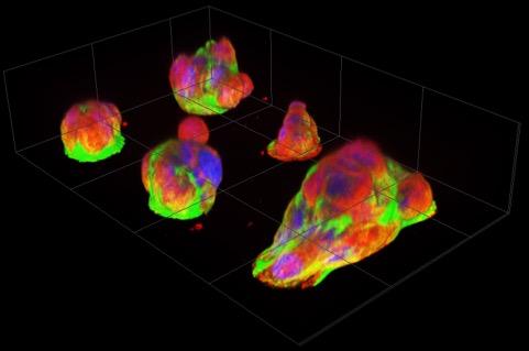
Image: Fluorescence images of pancreatic cancer microtumors after overnight culturing. Papillary structures pile up on micro-attachment sites (diameter 30 μm), with numerous cells visible per patch. The rightmost microtumor has extended over two attachment sites. Nuclei, actin filaments, and microtubules are labeled with blue, green and red fluorescent markers respectively. Image courtesy of Miyatake Y. et al., Scientific Reports.