It has been almost 20 years since the human genome was first sequenced, but researchers still know little about how the genome is folded up and organized within cells. In a paper published today in the Journal of Cell Biology, researchers from the University of Illinois at Urbana-Champaign describe a new technique that can measure the position of every single gene in the nucleus to build a 3D picture of the genome's organization.
The location of gene—whether they are close to the edge or the center of the nucleus, for example—may have significant effects on their activity, and a gene's position may therefore change as a cell develops or becomes diseased. Researchers can examine the position of individual genes using a microscope, but determining the position of every gene at the same time is impossible this way.
The new technique, called tyramide signal amplification sequencing (TSA-Seq), involves targeting an enzyme, horseradish peroxidase, to particular nuclear structures, such as the nuclear lamina that surrounds the nucleus or protein-containing granules called nuclear speckles that tend to be found in the center of the nucleus. The horseradish peroxidase then generates a highly reactive molecule called tyramide that can be used to label any DNA in the enzyme's vicinity. The closer a gene is to the enzyme, the more it will be labeled. So, when researchers subsequently sequence the cells' DNA, they can calculate how close each gene was to the nuclear structure tagged with horseradish peroxidase.
"TSA-Seq is the first genome-wide method capable of estimating actual distances of genes from particular nuclear subcompartments," says Yu Chen, first author on the paper.
Chen and colleagues tested their approach in leukemia cells and found that genes closer to nuclear speckles tended to be more active than genes closer to the nuclear lamina. Indeed, by examining the position of neighboring genes, the researchers were able to trace whole sections of chromosomes that looped out from the nuclear periphery toward speckles in the center of the nucleus. The function of nuclear speckles is unknown, but the regions of chromosomes close to speckles seem to be "hot zones" of gene activity.
"The logic of this nuclear organization remains to be determined, but our model would suggest that chromosome movements of just a few hundred nanometers could have substantial functional significance," Belmont says. A small shift in a gene's position so that it lies close to a nuclear speckle could be sufficient to dramatically enhance the gene's activity, for example.
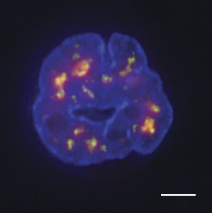
Image: Targeting horseradish peroxidase to nuclear speckles (green) produces a diffuse cloud of tyramide-containing molecules (red) that can label nearby DNA (blue). Sequencing the DNA can produce a map of the genome's organization. Image courtesy of Chen et al., 2018.