A team led by Purdue University has developed an improved three-dimensional super-resolution "nanoscope" for imaging key brain structures in great detail. Able to reconstruct a 3D images of 30-μm-thick brain sections, the nanoscope may help reveal how neurodegenerative diseases—such as Alzheimer's—begin.
 Single-molecule switching nanoscopy (SMSN) was first reported in Cell in 2016 by Fang Huang and others. The high-resolution method allowed for 3D imaging of cells as thick as 10 micrometers. However, while effective for visualizing cells, bacteria, and viruses in fine detail, the system lacks the power to accommodate naturally thicker samples, such as brain tissue.
"Brain tissue is particularly challenging for single molecule super-resolution imaging because it is highly packed with extracellular and intracellular constituents, which distort and scatter light - our source of molecular information. You can image deep into the tissue, but the image is blurry," said senior co-author Fang Huang, now an assistant professor at Purdue.
To overcome these limitations, the team developed new techniques for adjusting the optical mirrors in response to the tissue sample depths, compensating for light distortions introduced by the tissue. In addition, the technique also intentionally introduced extra aberrations to maintain the position information carried by a single molecule.
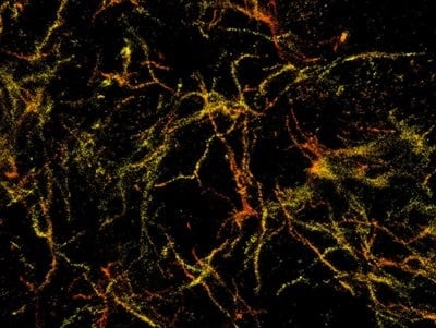
As reported today in Nature Methods, the team implemented this system on a frontal cortex section from a mouse model for Alzheimer's disease. They were to successfully reconstruct the brain’s whole tissue, its cells, and even the cell constituents. A Youtube video highlights the process.
“We combined active shaping of point spread functions and efficient adaptive optics to enable robust 3D-SMSN imaging within tissues. This development allowed us to image through 30-μm-thick brain sections to visualize and reconstruct the morphology and the nanoscale details of amyloid-β filaments in a mouse model of Alzheimer’s disease,” the team reported.
"While strictly a research tool for the foreseeable future, this technology has allowed us to see how the plaques are assembled and remodeled during the disease process. It gives insight into the biological causes of the disease, so that we can see if we can stop the formation of these damaging structures in the brain," said senior co-author Gary Landreth.
Image: A 3D single molecule super-resolution image of the amyloid plaques associated with Alzheimer's disease in 30-micron thick sections of the mouse's frontal cortex.