A new type of zebrafish that produces fluorescent tags in migratory embryonic nerve precursor cells has been created. The transgenic zebrafish line, in which the Fucci system is specifically expressed in delaminating and migrating neural crest cells, is described in a paper published last month in Genesis.
The zebrafish line produces fluorescent tags that glow brightly in different colors based on the behavior of a gene called SOX10 that is active in neural crest cells, transient embryonic stem cells that give rise to many cell types in the body, including neurons and cartilage. The cells can be seen transitioning in this time-lapse movie.
Why the cells stop dividing is one of the key questions that Rosa Uribe assistant professor of biosciences at Rice hopes to answer. SOX10 is one of more than 20 varieties of SOX proteins, and all of them regulate rapid cell division in fast-growing embryos. The same SOX proteins are also often found activated in cancer cells. Finding the "off" switch for SOX10 in neural crest cells could potentially lead to treatments for cancers where SOX proteins play a role, she said.
"Neural crest cells are stem cells that form from the earliest portion of our central nervous system, the neural tube," said Uribe. "They express SOX10 in addition to a bunch of other really important genes." To view neural crest cells during embryonic development, Uribe's team keeps a stock of breeder fish in a state-of-the-art fish room with hundreds of tanks—for the new reporter line and more than a dozen others—are removed from the tanks by hand each day and brought to the laboratory for observation.
Using a variety of methods, Uribe and her students immobilize live embryos and take photographs to trace their development over a period of hours. For example, the neural crest cells, which first appear in zebrafish embryos about 12 hours after fertilization, were tracked and observed for up to four hours. "Neural crest cells also do something else that's relevant to cancer," Uribe said. "They undergo something called the epithelial-to-mesenchymal transition, or EMT, shortly after they form, and this is what allows them to break away and migrate to the various places in the embryo."
Having the ability to observe neural crest cells from the moment they form until they finish migrating is one key to understanding them. Uribe's team will use the new cell line for this, in addition to others that have different colored tags for different reporter genes. They'll also mix and match genes in new strains of zebrafish to test what happens when cells make either too much of a specific protein or too little.
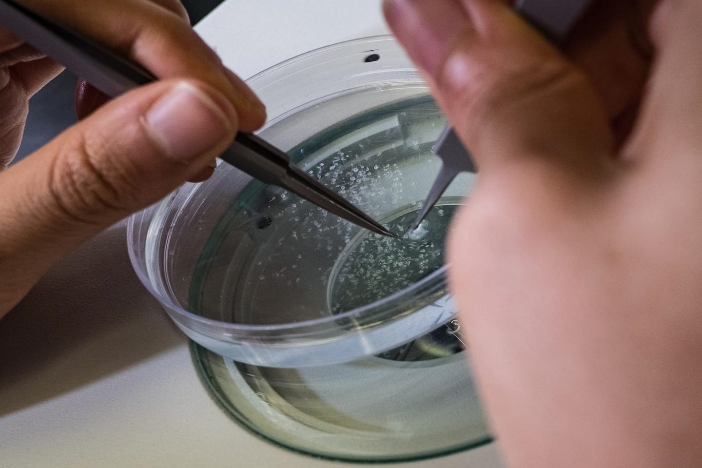
Image: Sorting zebrafish embryos in the Uribe lab at Rice University. Image courtesy of Jeff Fitlow, Rice University