Invadosomes are cellular structures observed in both normal cells and cancer cells. Their defining feature is the ability to interact with and degrade the extracellular matrix and is believed to be key structures of cell invasion. Previous in vivo studies have linked invadosomes to invasiveness and metastasis formation in cancer. Using a new method that combines laser dissection and proteomics, a team from Université de Bordeaux in France uncovers new insight in the formation of invadosomes.
 “The specific analysis of invadosomes is challenging because it is difficult to maintain their integrity during isolation. In addition, classical purification methods often suffer from contaminations, which may impair data validation,” the team notes in their recent publication in Nature Communications.
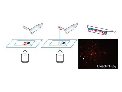
The team generated an invadosome cell culture model that were transfected with a red fluorescent marker. This allowed for the detection of invadosomes without exogenous staining. Then, by using an automated laser microdissector, selected regions were cut and propelled onto the cap of a small microcentrifuge tube. Cells imaged before and after microdissection confirmed proper collection of invadosomes.
After collection of 40,000 invadosome particles, or rosettes, the team extracted proteins and analyzed them by liquid chromatography-tandem MS (LC-MS/MS). The team identified 312 proteins that were exclusively enriched in the invadosomes. Surprisingly a large number of these proteins were found to be involved in mRNA translation. Treatment with translation inhibitors anisomycin and cycloheximide impacted invadosome formation, confirming the role of an associated translational machinery.
The team concludes that protein translation activity is an inherent property of invadosomes, which helps control their formation.
“Translational control is also crucial in cancer development, especially the selective control of the translation of specific mRNAs that promote tumor progression including invasion and metastasis,” the team adds.
Image: A schematic representation of the microdissection process. The laser cuts the region of interest, which will then be propelled into the cap of a tube. Image courtesy of Ezzoukhry et al. and Nature Communications.